Chart to Compare Three Types of Muscles, taken from "Essentials of Anatomy and Physiology"
| Cardiac Muscle | Skeletal Muscle | Smooth Muscle | |
| Location | Heart | Attached to bone | On hollow organs, glands and blood vessels |
| Cell Shape | Branched | Cylnidrical | Spindle-shaped |
| Nucleus | Central & single | Multiple, peripheral | Single, central |
| Special Features | Intercalated disks | Cell-Cell attachment | |
| Striations | yes | yes | no |
| Autorhythmic | yes | no | yes |
| Control | involuntary | voluntary | involuntary |
| Function | Move the whole body | Heart contraction to propel blood | Compression of ducts and tubes, etc |
SKELETAL SYSTEM
The human body contains 206 bones. The skeletal system consists of bone, cartilage and connective tissue. The connective tissue is a matrix and the cells that produce it, there is a variety of amounts of collagen, proteoglycan and minerals in the matrix that determine exactly what type of matrix it is.
The bone has two major types: compact and cancellous. Compact bone has haversian systems which consist of osteocytes organized into lamellae surrounding haversian canals. Cancellous bone tissue has trabeculae without the haversian canals. Bones grow, remodel and repair during a persons lifetime. The growth of the bones is either on the surface of existing bone or occurs within the cartilage. Bone remodeling is when there is a removal of old bone material by osteoclasts and new bone material is replaced by osteoblasts. When a bone is broken, it repairs itself by cells moving into the damaged area to form a callus, which eventually is replaced by bone.
There are two major skeletal breakdowns in the human body, the axial skeleton and appendicular skeleton. The axial skeleton consists of the skull, vertebral column and thoracic cage. The appendicular skeleton consists of every other bone, for example the limbs and their girdles. Along with these breakdowns of the system, the skeletal system is also has articulations, also known as joints. There are three major types of joints: fibrous, cartilaginous and synovial. Fibrous joints are joined by fibrous connective tissue and while one would usually characterize joints with movement, this type of joint allows little or no movement. Cartilaginous joints are united by cartilage and allow some movement. Synovial joints are the major joints such as plane, saddle, hinge, pivot, etc and allow the most movement. To find more information on Synovial Joints, see the diagrams below.
How The Systems Interact
The skeletal system and the muscular system work together to allow movement throughout the body. Without this unison, no one would be able to move or conduct what are considered simple and everyday tasks.
The skeletal system is made up of bones and their associated cartilage as well as joints, which allow movement. The main function of the skeletal system is to protect, support and allow body movement. Also, the skeletal system produces blood cells and stores minerals. The muscular system is made up of muscles that are attached to the skeleton. There are three different types of muscle, each with their own specific function. The three types are: cardiac, skeletal and smooth muscle. The main function of the muscle system is to allow body movement, maintain posture and produce body heat.
The skeletal and muscular system must work together to allow movement. Joints are considered part of the skeletal system and joints allow movement through a variety of types of joints. However, these joints can�t move on their own. They need muscles to allow movement. Also, most of the muscles in the body, especially skeletal muscles, have an �origin� on one bone and an �insertion� onto another bone. They also usually cross at least one joint.
These systems are key to the body�s ability to function. The skeleton provides the structure of the human body, but the muscles allow the skeleton to carry out the tasks necessary in day-to-day life.
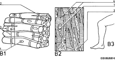
Skeletal Muscle
B1 Skeletal (striated) muscle
1 Muscle cell nuclei
2 Connective tissue
3 Striations
B2 Longitudinal section
1 Muscle cell nuclei
2 Connective tissue
4 Intercalated discs
B3 Typical location
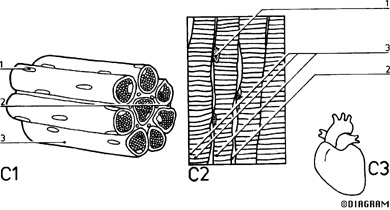
Cardiac Muscle
C1 Cardiac muscle
1 Muscle cell nuclei
2 Connective tissue
3 Striations
C2 Longitudinal section
1 Muscle cell nuclei
2 Connective tissue
3 Striations
C3 Location
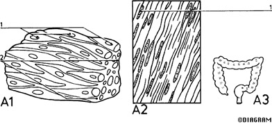
Smooth Muscle
A1 Smooth muscle
1 Muscle cell nuclei
2 Connective tissue
A2 Longitudinal section
1 Muscle cell nuclei
A3 Typical location
SKELETAL SYSTEM DIAGRAMS
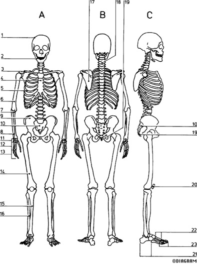
Skeletal System, Anterior, Posterior and Lateral View
A Anterior view
B Posterior view
C Lateral view
1 Cranium 2 Mandible
3 Clavicle 4 Sternum
5 Ribs 6 Humerus
7 Radius 8 Ulna
9 Pelvic girdle 10 Carpus
11 Metacarpus 12 Phalanges
13 Femur 14 Tibia
15 Fibula 16 Scapula
17 Spinal vertebrae 18 Patella
19 Tarsus 20 Metatarsus
21 Phalanges
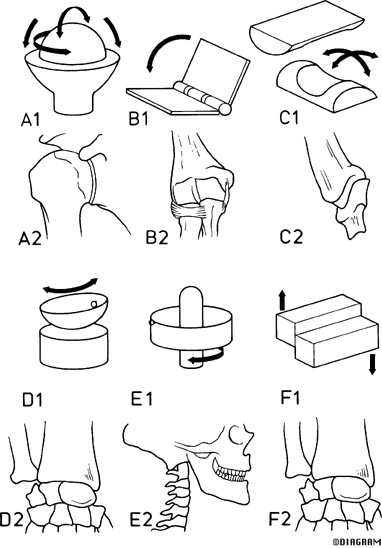
Types of Synovial Joints
A1 Ball-and-socket (spheroidal) joint mechanism A2 Shoulder joint
B1 Hinge joint (ginglymus) mechanism B2 Elbow joint
C1 Saddle (sellar) joint mechanism C2 Carpometacarpal joint of thumb
D1 Ellipsoid joint mechanism D2 Wrist (radiocarpal) joint
E1 Pivot (trochoid) joint mechanism E2 Median atlanto-axial joint
F1 Plane (gliding) joint mechanism F2 Intercarpal joints
LINKS
Muscular System Links:
Human Muscular System
The Hosford Muscle Tables:
Skeletal Muscles of the Human Body
WebAnatomy: Muscular System
Skeletal System Links:
Human Anatomy Online: Skeletal System
Gray's Anatomy of the Human Body: Osteology
Illustrated Encyclopedia of Human Anatomic Variation: Skeletal System
Miscellaneous Links:
Human Anatomy Diagrams
Images/Diagrams taken from Human Anatomy Diagrams
Other information gathered from links above and textbook, "The Essentials of Anatomy and Phsyiology"
This website was created by Rachel Johnson on June 3, 2003 for the final exam in Mrs. Edwards Human Anatomy Class.