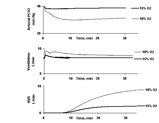
Fig. 6. Arterial PCO2, ventilation and hypoxic ventilatory depression while breathing 10% and 15% O2, during quiet wakefulness. Ventilation peaks almost immediately after the disturbance then declines, as HVD develops, to a steady state value. Arterial PCO2 declines to a minimum then rises to a steady state value, mirroring the pattern of ventilation.