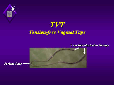
The instrument consists of two needles to which a prolene tape covered by a plastic sheath is connected.
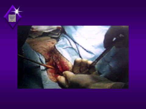
Through a small vaginal incision (1.5 cm) one needle is introduced behind the symphysis pubis on one side of the mid-urethra.
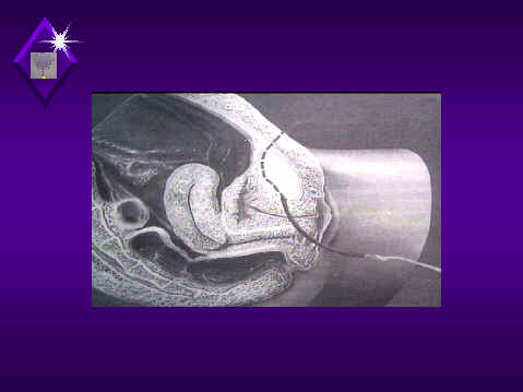
A diagrammatic illustration of insertion of the needle behind the symphysis pubis and is brought up through the abdominal wall.
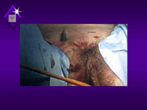
The needle is brought up through the abdominal wall above the symphysis pubis through 1 cm incision.
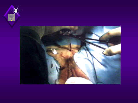
The same procedure is repeated with the other needle which is inserted along the other side of the mid-urethra. The two needles are then cut
The tape is adjusted under the mid-urethra to avoid incontinence.
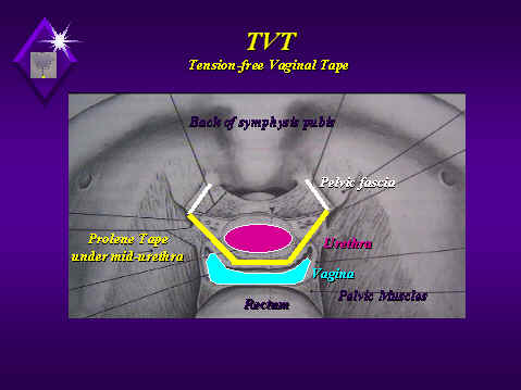
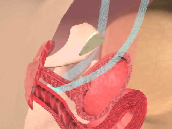
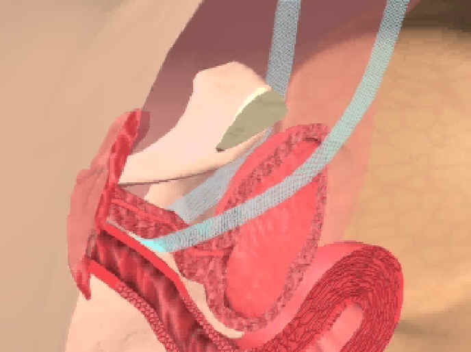
A diagrammatic presentation of the location of the tape (yellow) inserted under mid-urethra (dark pink) to support the urethra in the same way the pubo-urethral ligaments normally support the urethra.
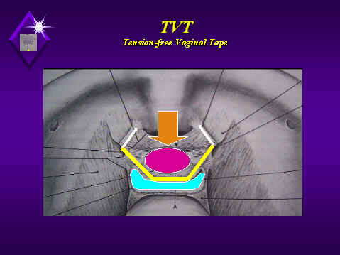
The tape in position to support the urethra helping its occlusion during the increase of intra-abdominal pressure (orange arrow).
During absence of physical effort:
The tape in position under the mid urethra without any tension

During physical effort (e.g. coughing):
The tape in position to support the urethra helping its occlusion during the increase of intra-abdominal pressure (The urethra become occluded against the Tape)

The previous 2 slides together for easy comparison of the position of the urethra during rest and physical activity