

PATHOLOGICAL VARIATIONS ENDOSCOPICALLY DETECTED
VARIANTI PATOLOGICHE RILEVATE IN ENDOSCOPIA
torna alla
Malformations
|
|
|
|
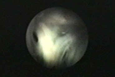
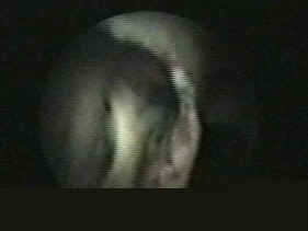
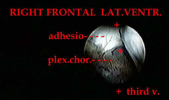
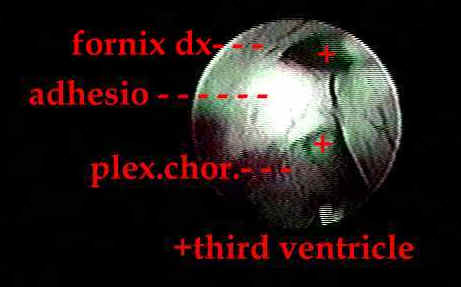
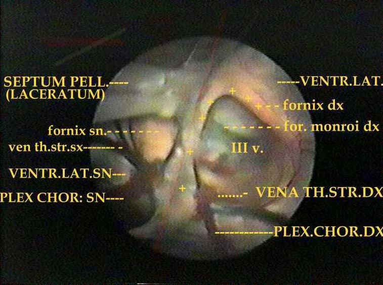
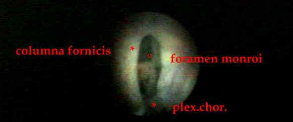
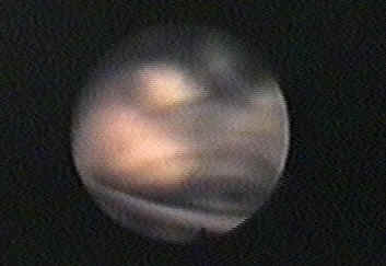
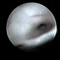
This picture shows the appearance of the monroi foramen in a patient affected by myelomeningocoele.The foramen seems divided in two partand You could be disoriented in defining which is the correct way to reach the third ventricle...
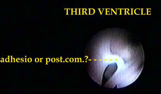
This particular aspect is due to an exceedingly high adhesio which actually divides the monroi foramen in a posterior part with the choroidal plexus and an anterior part without them...
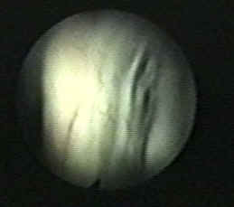
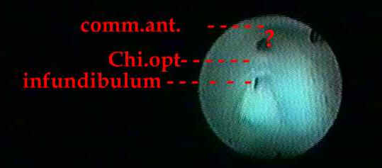
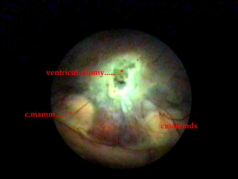
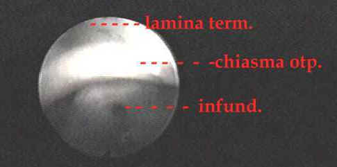
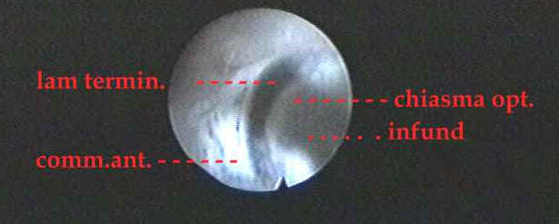
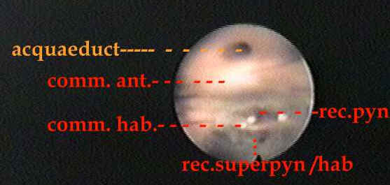
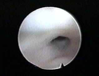
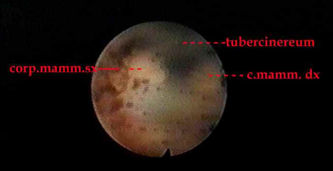
Hydrocephalus in myelomeningocoele: the anterior part of the third ventricle seen from above and behind . This is a good example of how advantageous is an extensive knowledge othe normal anatomy!
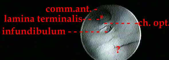

Hydrocephalus in myelomeningocoele
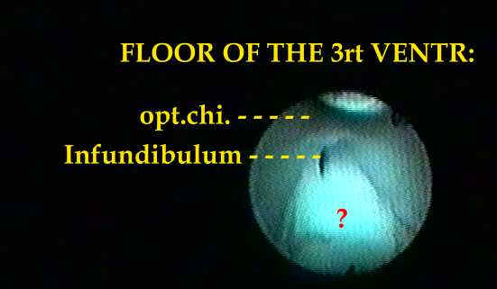
malformed part of the anterior third ventricle. Mammillary bodies can not be identified

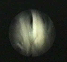
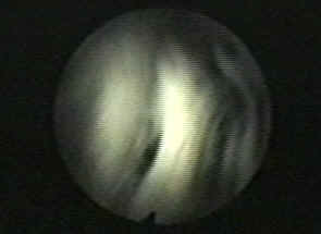
Just anteriorly to the mammillary body the apex of the basilar artery (or P1) may herniate through the attenuated floor of the third ventricle
Long lasting hydrocephalic dilatation
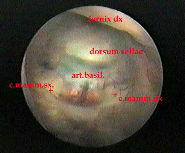
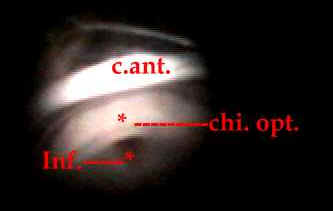
inf. infundibulum
tub. cin.tuber cinereum
a.b. arteria basilaris (translucent)
p1. arteria cerebri posterior


idem
idem particolare
idem particolare
idem particolare

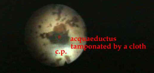
Intraventricular hemorrhages
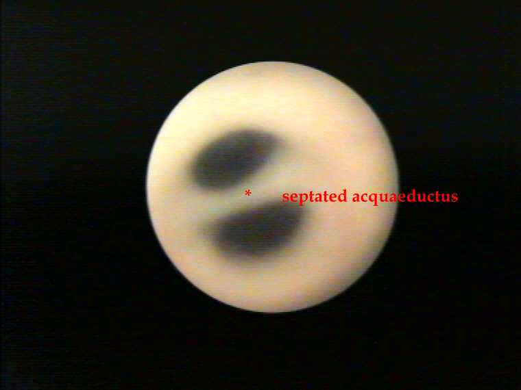
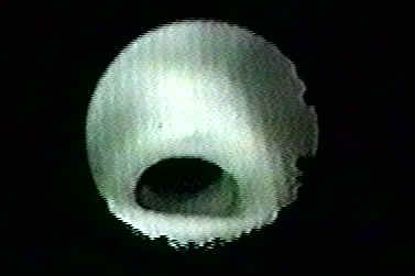
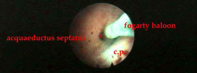
2months infant with post hemorrhagic hydrocephalus and trapped fourth ventricle
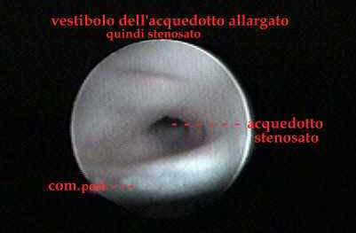
The cloth obstructing the acquaeduct is removed, but underneath a membrane has developped and is perforated
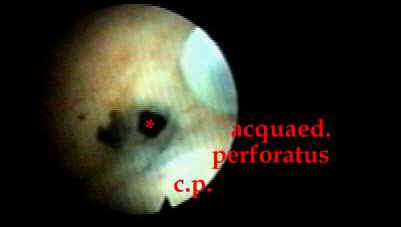
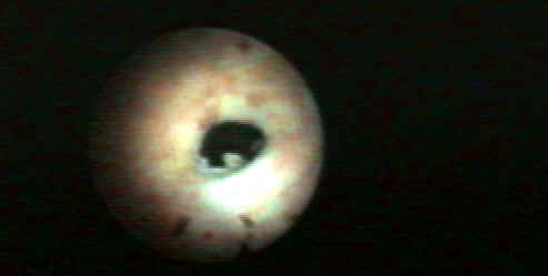
final result after acquaeductoplasty
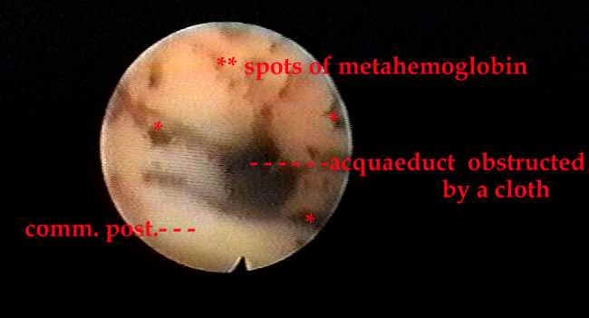
Posthemorrhagic modifications
of the internal surface of the
ventricles are characteristic and very common in paediatric
patients. The ependimal layer reacts to the deposit of small
particles of metahemoglobin with the production of a protective
membrane. The nature of which is quite similar to the structure of the
arachnoid. So upon the inner surface of the lateral ventricles a
web is stretched which hides quite every structure and
the foramen of MONRO itself. Yet if this membrane is teased
a little bit, you will find the normal ependimal laye. If csf flows between these layers
and ependyma it can accumulate and it can cause
a cyst. Many of these enlarged cysts cause the so-called
pluriseptaded hydrocephalus;
2months olf infant with posthemorrhagic hydrocephalus and trapped fourth ventricle
Posthemorrhagic hydrocephalus changes the normal anatomy of the cerebral
ventricles, the scene become unfamiliar and easily one
can get lost. When Choroid plexus are concealed
by membranes the optostriate sulcus andparticularly
the convergence of the three main vein ( thalamostriate, septal, caudate) toward the foramen of monro can be of great
valuein orientingthe neurosurgeon