Orthopedic Surgery
Each pelvic (hip) radiograph submitted to the OFA is evaluated by three diplomates of the American College of Veterinary Radiologists. By consensus the hip joint conformation is classified as excellent, good, fair, borderline, mildly dysplastic or severely dysplastic. Classification as excellent, good or fair is considered within normal limits. If the dog is two years of age or older, a breed registry number is assigned, and the dog is then certified. Dogs must be two years of age because it has been established that 94% of all dysplastic dogs will show radiographic evidence of their disease by two years of age.
The ultimate purpose of OFA certification is to provide information to dog owners to assist them with the selection of good breeding animals. Therefore, attempts to get a dysplastic dog certified will only hurt the breed by perpetuation of the disease.
PennHIP (University of Pennsylvania Hip Improvement Program) incorporates a new method for evaluating the integrity of the canine hip. It is accurate in puppies as young as 16 weeks of age. It has great potential to lower the frequency of CHD when used as a selection criterion.
PennHIP is a multifaceted radiographic technology (x-ray) for hip evaluation. The technique assesses the quality of the canine hip and quantitatively measures canine hip joint laxity. The PennHIP method of evaluation is more accurate than the current standard in its ability to predict the onset of osteoarthritis. Osteoarthritis, also known as degenerative joint disease (DJD), is the hallmark of canine hip dysplasia (CHD).
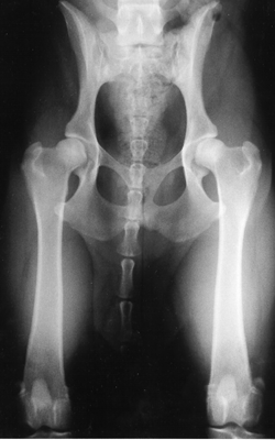
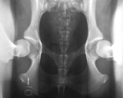
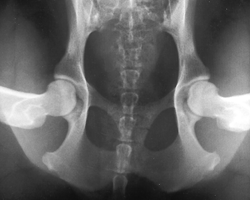
The PennHIP method is a different way to assess, measure and interpret hip joint laxity. It consists of three separate radiographs: the distraction view, the compression view and the hip-extended view (see below). The distraction view and compression view, developed by Dr. Smith, are used to obtain accurate and precise measurements of joint laxity and congruity. The hip-extended view is used to obtain supplementary information regarding the existence of DJD in the hip joint.