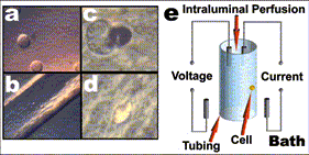

Click image to go to larger resolution.
Fig. 3.
Tubing system for simultaneous single cell
voltage-clamp and intracellular perfusion. (a-d) Images taken with a custom inverted microscope (Bustamante,
1991). The images show the process of
forming a seal between the hole in the plastic pipette (e.g. Bustamante &
McDonald, 1983) and a neuroblastoma cell (about 20 mm in
diameter). (a) Neuroblastoma
cells. (b) Low magnification view of
the tubing placed over the target cell.
(c) High magnification view of the tubing placed just over the target
cell, a few seconds prior applying a gentle suction to attract the cell towards
the hole in the tube. (d) High
magnification view of the cell in the hole ready for voltage-clamp and
intracellular perfusion (achieved after a vacuum pulse). From Bustamante (1983). (e) Diagram showing the placement of the two
pairs of half-cells, Ag/AgCl electrodes, for the simultaneous recording of
current and voltage. The 4-electrode
system is a requirement of experimental arrangements where large polarizing
currents are used (e.g. Hodgkin and Huxley, 1952). In the voltage-clamp configuration, the detection system
incorporates a circuit for the compensation of the resistance in series to the
target, Rseries (e.g. Bustamante and McDonald, 1983).