#
Assignment 3:
Visualization
of 3D Volumetric Structures from Medical Images
|
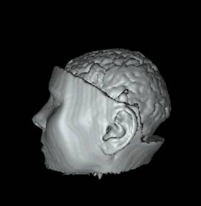
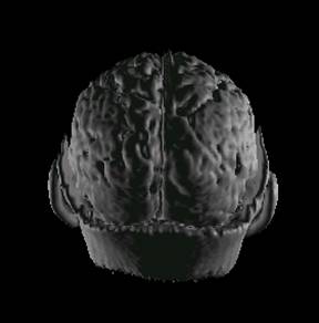
Algorithme Used
: RAY TRACING |
|
|
Software Used : VOLVIS |
File
Type : Slice File Data
Size : 128 x 128 x 84 voxels Voxel Size : 1.000000 x 1.000000 x 1.000000 mm 8 bits per voxel Data
Origin : Magnetic Resonance Data |
|
Description : The image is constructed
by using Volvis software. The file is a slice file type
of human head and brain. It can be rendered by volvis
and controlled by adjusting position view, color, background, speed etc. Below is the two result of rendering by
using ray tracing method. |
|
|
Snapshot : see from side |
Snapshot : see in backward |
|
Animation link : http://www-sop.inria.fr/epidaure/personnel/malandain/malandain_segment.html |
|
|
|
|
|
Algorithme Used
: MARCHING CUBES |
|
|
Software Used : VOLVIS |
File
Type : Slice File Data
Size : 128 x 128 x 84 voxels Voxel Size : 1.000000 x 1.000000 x 1.000000 mm 8 bits per voxel Data
Origin : Magnetic Resonance Data |
|
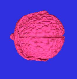
Description : The image is constructed
by using Volvis software. The file is a slice file
type of human head and brain. The two images below is rendered by volvis and controlled by adjusting position view, color,
background, speed etc. Below is the
two result of rendering by using marching cubes methods. |
|
|
Snapshot : see at x axis |
snapshot : in z direction |
|
Links to more medical images: http://www.barre.nom.fr/medical/samples/ ftp://ftp-wjq.philips.com/medical/interoperability/out/Medical_Images/ http://www.comp.leeds.ac.uk/comir/resources/links_c.html http://www.erl.wustl.edu/DICOM/96_image_report_index.html ftp://imrad.ucdmc.ucdavis.edu/pub/dicom/UCDMC/GE_MRI_CT_TestFiles/ ftp://wuerlim.wustl.edu/pub/dicom/images/version3/ |
|
|
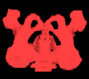
Algorithme Used
: Maximum Intensity
Projection |
|
|
Software Used : 3 D DOCTOR |
File type and size :
unknown Resources : 3 D Doctor
software |
|
Description : The image is constructed
by using 3 D Doctor software, using volume rendering and maximum density type
with 1/1 resolution. |
|
|
Image : Pelvis
|
|
|
Other information : http://www.vaytek.com/booksD.html http://www.medicimaging.com/medicview/Demo/Download.htm |
|