Image courtesy of MedValet
The Heart and the Circulatory System
Types of Circulatory Systems
Remember that the circulation of the blood serves to move blood to a site or sites where it can be oxygenated, and where wastes can be disposed. Circulation then serves to bring newly oxygenated blood to the tissues of the body. As oxygen and other chemicals diffuse out of the blood cells and into the fluid surrounding the cells of the body's tissues, waste produces diffuse into the blood cells to be carried away. Blood circulates through organs such as the liver and kidneys where wastes are removed, and back to the lungs for a fresh dose of oxygen. And then the process repeats itself. This process of circulation is necessary for continued life of the cells, tissues and even of the whole organisms. Before we talk about the heart, we should give a brief background of the two broad types of circulation found in animals. We will also discuss the progressive complexity of the heart as one moves up the evolutionary ladder.
Many invertebrates do not have a circulatory system at all. Their cells are close enough to their environment for oxygen, other gases, nutrients, and waste products to simply diffuse out of and into their cells. In animals with multiple layers of cells, especially land animals, this will not work, as their cells are too far from the external environment for simple osmosis and diffusion to function quickly enough in exchanging cellular wastes and needed material with the environment.
Open Circulatory Systems
In higher animals, there are two primary types of circulatory systems -- open and closed. Arthropods and most mollusks have an open circulatory system. In this type of system, there is neither a true heart or capillaries as are found in humans. Instead of a heart there are blood vessels that act as pumps to force the blood along. Instead of capillaries, blood vessels join directly with open sinuses. "Blood," actually a combination of blood and interstitial fluid called 'hemolymph', is forced from the blood vessels into large sinuses, where it actually baths the internal organs. Other vessels receive blood forced from these sinuses and conduct it back to the pumping vessels. It helps to imagine a bucket with two hoses coming out of it, these hoses connected to a squeeze bulb. As the bulb is squeezed, it forces the water along to the bucket. One hose will be shooting water into the bucket, the other is sucking water out of the bucket. Needless to say, this is a very inefficient system. Insects can get by with this type system because they have numerous openings in their bodies (spiracles) that allow the "blood" to come into contact with air.
Closed Circulatory Systems
The closed circulatory system of some mollusks and all higher invertebrates and the vertebrates is a much more efficient system. Here blood is pumped through a closed system of arteries, veins, and capillaries. Capillaries surround the organs, making sure that all cells have an equal opportunity for nourishment and removal of their waste products. However, even closed circulatory systems differ as we move further up the evolutionary tree.
One of the simplest types of closed circulatory systems is found in annelids such as the earthworm. Earthworms have two main blood vessels -- a dorsal and a ventral vessel -- which carry blood towards the head or the tail, respectively. Blood is moved along the dorsal vessel by waves of contraction in the wall of the vessel. These contractible waves are called 'peristalsis.' In the anterior region of the worm, there are five pairs of vessels, which we loosely term "hearts," that connect the dorsal and the ventral vessels. These connecting vessels function as rudimentary hearts and force the blood into the ventral vessel. Since the outer covering (the epidermis) of the earthworm is so thin and is constantly moist, there is ample opportunity for exchange of gases, making this relatively inefficient system possible. There are also special organs in the earthworm for the removal of nitrogenous wastes. Still, blood can flow backward and the system is only slightly more efficient than the open system of insects.
As we come to the vertebrates, we begin to find real efficiencies with the closed system. Fish possess one of the simplest types of true heart. A fish's heart is a two-chambered organ composed of one atrium and one ventricle. The heart has muscular walls and a valve between its chambers. Blood is pumped from the heart to the gills, where it receives oxygen and gets rid of carbon dioxide. Blood then moves on to the organs of the body, where nutrients, gases, and wastes are exchanged. However, there is no division of the circulation between the respiratory organs and the rest of the body. That is, the blood travels in a circuit which takes blood from heart to gills to organs and back to the heart to start its circuitous journey again.
Frogs have a three-chambered heart, consisting of two atria and a single ventricle. Blood leaving the ventricle passes into a forked aorta, where the blood has an equal opportunity to travel through a circuit of vessels leading to the lungs or a circuit leading to the other organs. Blood returning to the heart from the lungs passes into one atrium, while blood returning from the rest of the body passes into the other. Both atria empty into the single ventricle. While this makes sure that some blood always passes to the lungs and then back to the heart, the mixing of oxygenated and deoxygenated blood in the single ventricle means the organs are not getting blood saturated with oxygen. Still, for a cold-blooded creature like the frog, the system works well. Humans and all other mammals, as well as birds, have a four-chambered heart with two atria and two ventricles. Deoxygenated and oxygenated blood are not mixed. The four chambers ensure efficient and rapid movement of highly oxygenated blood to the organs of the body. This has helped in thermal regulation and in rapid, sustained muscle movements.
We have learned much about the heart and circulatory system since Harvey's pioneering work. Scientific research has replaced mystical spirits as the basis for medical practice. In the next part of this chapter, thanks to the work of William Harvey, we will discuss our human heart and circulation, some of the medical problems that can occur, and how advances in modern medical care allow treatment of some of these problems.
Heart Chambers
|
Image courtesy of MedValet |
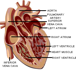
There are four chambers in the heart - two atria and two ventricles. The atria (one is called an atrium) are responsible for receiving blood from the veins leading to the heart. When they contract, they pump blood into the ventricles. However, the atria do not really have to work that hard. Most of the blood in the atria will flow into the ventricles even if the atria fail to contract. It is the ventricles that are the real workhorses, for they must force the blood away from the heart with sufficient power to push the blood all the way back to the heart (this is where the property of contracting with more force when stretched comes into play). The muscle in the walls of the ventricles is much thicker than the atria. The walls of the heart are really several spirally wrapped muscle layers. This spiral arrangement results in the blood being wrung from the ventricles during contraction. Between the atria and the ventricles are valves, overlapping layers of tissue that allow blood to flow only in one direction. Valves are also present between the ventricles and the vessels leading from it.
Cardiac Conduction
Though the brain can cause the heart to speed up or slow drain, it does not control the regular beating of the heart. As noted earlier, the heart is composed of involuntary muscle. The muscle fibers of the heart are also self-excitatory. This means they can initiate contraction themselves without receiving signals from the brain. This has been demonstrated many times in high school classes of the past by removing the heart of a frog or turtle, and then stimulating it to contract. The heart continues to beat with no further outside stimulus, sometimes for hours if bathed in the proper solution. In addition, cardiac muscle fibers also contract for a longer period of time than do skeletal muscles. This longer period of contraction gives the blood time to flow out of the heart chambers.
The heart has two areas that initiate impulses, the SA or sinoatrial node, and the AV or atrioventricular node. The heart also has special muscle fibers called Purkinje fibers that conduct impulses five times more rapidly than surrounding cells. The Purkinje fibers form a pathway for conduction of the impulse that ensures that the heart muscle cells contract in the most efficient pattern. The SA node is located in the wall of the right atrium, near the junction of the atrium and the superior vena cava. This special region of cardiac muscle contracts on its own about 72 times per minute. In contrast, the muscle in the rest of the atrium contracts on its own only 40 or so times per minute. The muscle in the ventricles contracts on its own only 20 or so times per minute. Since the cells in the SA node contract the most times per minute, and because cardiac muscle cells are connected to each other by intercalated discs, the SA node is the pacemaker of the heart. When the SA node initiates a contraction, Purkinje fibers rapidly conduct the impulse to another site near the bottom of the right atrium and near the center of the heart. This region is the AV node, and slows the impulse briefly. The impulse then travels to a large bundle of Purkinje fibers called the Bundle of His, where they move quickly to the septum that divides the two ventricles. Here, the Purkinje fibers run in two pathways toward the posterior apex of the heart. At the apex, the paths turn in opposite directions, one running to the right ventricle, and one running to the left. The result is that while the atria are contracting, the impulse is carried quickly to the ventricles. With the AV node holding up the impulse just enough to let the atria finish their contraction before the ventricles begin to contract, blood can fill the ventricles. And, since the Purkinje fibers have carried the impulse to the apex of the ventricles first, the contraction proceeds from the bottom of the ventricles to the top where the blood leaves the ventricles through the pulmonary arteries and the aorta.
Heart Sounds
The contraction of the heart and its anatomy cause the distinctive sounds heard when listening to the heart with a stethoscope. The "lub-dub" sound is the sound of the valves in the heart closing. When the atria end their contraction and the ventricles begin to contract, the blood is forced back against the valves between the atria and the ventricles, causing the valves to close. This is the "lub" sound, and signals the beginning of ventricular contraction , known as systole. The "dub" is the sound of the valves closing between the ventricles and their arteries, and signals the beginning of ventricular relaxation, known as diastole. A physician listening carefully to the heart can detect if the valves are closing completely or not. Instead of a distinctive valve sound, the physician may hear a swishing sound if they are letting blood flow backward. When the swishing is heard tells the physician where the leaky valve is located.
The Pulmonary and Systemic Circuits
The
Pulmonary Circuits
The heart is responsible for pumping the blood to every cell in the body. It is also responsible for pumping blood to the lungs, where the blood gives up carbon dioxide and takes on oxygen. The heart is able to pump blood to both regions efficiently because there are really two separate circulatory circuits with the heart as the common link. Some authors even refer to the heart as two separate hearts--a right heart in the pulmonary circuit and left heart in the systemic circuit. In the pulmonary circuit, blood leaves the heart through the pulmonary arteries, goes to the lungs, and returns to the heart through the pulmonary veins.
The Systemic Circuits
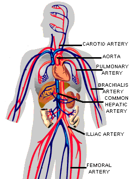
In the systemic circuit, blood leaves the heart through the aorta, goes to all the organs of the body through the systemic arteries, and then returns to the heart through the systemic veins. Thus there are two circuits. Arteries always carry blood away from the heart and veins always carry blood toward the heart. Most of the time, arteries carry oxygenated blood and veins carry deoxygenated blood. There are exceptions. The pulmonary arteries leaving the right ventricle for the lungs carry deoxygenated blood and the pulmonary veins carry oxygenated blood. If you are confused as to which way the blood flows through the heart, try this saying "When it leaves the right, it comes right back, but when it leaves the left, it's left." The blood does not have to travel as far when going from the heart to the lungs as it does from the heart to the toes. It makes sense that the heart would be larger on one side than on the other. When you look at a heart, you see that the right side of the heart is distinctly smaller than the left side, and the left ventricle is the largest of the four chambers.
Blood Supply to the Heart
While you might think the heart would have no problem getting enough oxygen-rich blood, the heart is no different from any other organ. It must have its own source of oxygenated blood. The heart is supplied by its own set of blood vessels. These are the coronary arteries. There are two main ones with two major branches each. They arise from the aorta right after it leaves the heart. The coronary arteries eventually branch into capillary beds that course throughout the heart walls and supply the heart muscle with oxygenated blood. The coronary veins return blood from the heart muscle, but instead of emptying into another larger vein, they empty directly into the right atrium.
The Blood Vessels
Blood Vessel Anatomy
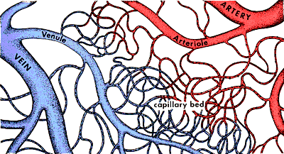
We need to briefly discuss the anatomy of the vessels. There are three types of vessels - arteries, veins, and capillaries. Arteries, veins, and capillaries are not anatomically the same. They are not just tubes through which the blood flows. Both arteries and veins have layers of smooth muscle surrounding them. Arteries have a much thicker layer, and many more elastic fibers as well. The largest artery, the aorta leaving the heart, also has cardiac muscle fibers in its walls for the first few inches of its length immediately leaving the heart. Arteries have to expand to accept the blood being forced into them from the heart, and then squeeze this blood on to the veins when the heart relaxes. Arteries have the property of elasticity, meaning that they can expand to accept a volume of blood, then contract and squeeze back to their original size after the pressure is released. A good way to think of them is like a balloon. When you blow into the balloon, it inflates to hold the air. When you release the opening, the balloon squeezes the air back out. It is the elasticity of the arteries that maintains the pressure on the blood when the heart relaxes, and keeps it flowing forward. if the arteries did not have this property, your blood pressure would be more like 120/0, instead of the 120/80 that is more normal. Arteries branch into arterioles as they get smaller. Arterioles eventually become capillaries, which are very thin and branching.
|
Image courtesy of Carolina Biological Supply/Access Excellence |
Capillaries are really more like a web than a branched tube. It is in the
capillaries that the exchange between the blood and the cells of the body takes
place. Here the blood gives up its carbon dioxide and takes on oxygen. In the
special capillaries of the kidneys, the blood gives up many waste products in
the formation of urine. Capillary beds are also the sites where white blood
cells are able to leave the blood and defend the body against harmful invaders.
Capillaries are so small that when you look at blood flowing through them under
a microscope, the cells have to pass through in single file. As the capillaries
begin to thicken and merge, they become venules. Venules eventually become veins
and head back to the heart. Veins do not have as many elastic fibers as
arteries. Veins do have valves, which keep the blood from pooling and flowing
back to the legs under the influence of gravity. When these valves break down,
as often happens in older or inactive people, the blood does flow back and pool
in the legs. The result is varicose veins, which often appear as large purplish
tubes in the lower legs.
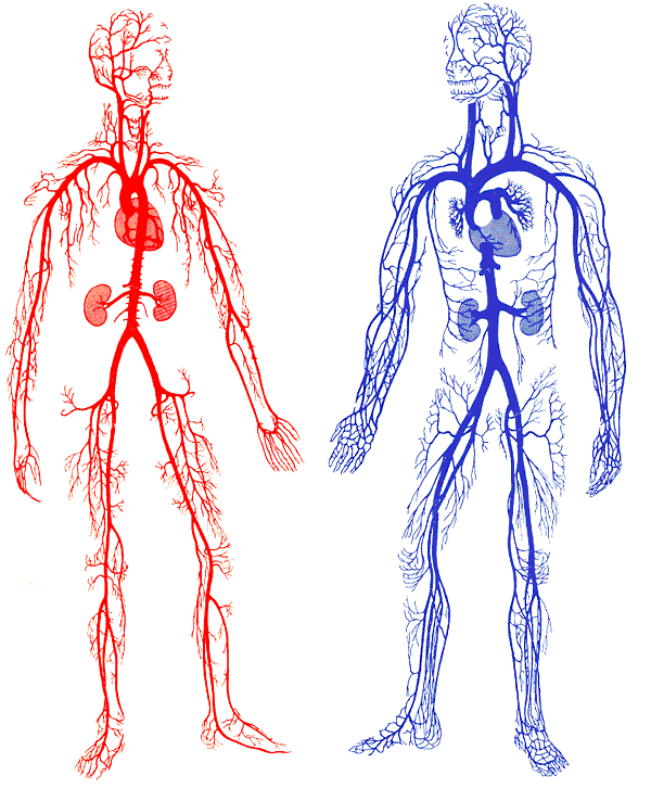
Arteries
|
Arteries in Red
Capillaries
|
What are capillaries?
Capillaries are extremely small vessels located within the tissues of the body that transport blood from the arteries to the veins. Capillary walls are thin and are composed of endothelium (a single layer of overlapping flat cells). Oxygen, carbon dioxide, nutrients and wastes are exchanged through the thin walls of the capillaries. The flow of blood is controlled by structures called precapillary sphincters. These structures are located between arterioles and capillaries and contain muscle fibers that allow them to contract. When the sphincters are open, blood flows freely to the capillary beds of body tissue. When the sphincters are closed, blood is not allowed to flow through the capillary beds. Capillary Size Capillaries are so small that red blood cells can only travel through them in single file.
|