________________________________________________ >>Difinition and Pathogenesis: Defined as a chronic disease of the liver in which widespread hepatic parenchymal cell destruction has led to the formation of connective tissue and nodular regeneration with consequent disorganization of the normal lobular architecture. * The liver nodules alter the anatomical location and function of the blood vessels and bile ducts, causing obstruction and resulting in portal hypertension, ascites, jaundice, hepatic encephalopathy and esophageal varices. * Pathogenetically, liver cirrhosis should be regarded as the final common pathway of chronic liver injury, which can result from any form of intense repeated prolonged liver cell injury. >>Major precipitating factors: Malnutrition and Genetic factors. >>Etiology and classification: >Etiology Cirrhosis is the terminal sequel of alcoholic liver disease or viral hepatitis, particularly Hepatitis C. Other, less frequent causes include some parasitic infections (such as Schistosomiasis), some metabolic disorders (such as Wilsons disease), toxic chemicals and unknown conditions. >Classification: 1. Alcoholic Cirrhosis (Laennec's): It has 2 actions: Direct and Indirect effect. - Direct: alcoholic abuse and biochemical alteration lead to oxidation of alcohol and so fatty changes (fatty infiltration) occur leading to inflammation and scarring liver. In the early stage, this fatty infiltration is not associated with fibrosis and scarring. Later stages are marked by a prominent inflammation, an increase in fibrous tissue, and a progressive shrinkage, nodularity, and hardening of the liver. - Indirect: induces the microsomal enzyme system resulting in highly toxic metabolites (ex. Paracetamol). 2. Biliary: Refers to cirrhosis following chronic obstruction of bile flow. - Primary biliary cirrhosis follows long-standing cholestasis that is generally of unknown etiology, but it may have an underlying immunologic basis with elevated IgM, Abs and circulating complement-fixing immune complexes. - Secondary biliary cirrhosis may be caused by stones or a tumor obstructing bile flow, leading to an inflammatory reaction and scarring. 3. Postnecrotic: scarring following massive hepatic necrosis such as that seen in chronic viral hepatitis, after exposure to hepatotoxic drugs, or in immunemediated hepatitis. 4. Metabolic: excessive iron (hemochromatosis) or copper (Wilson's disease) deposition, �-1-antitrypsin deficiency, other inborn errors of metabolism. 5. Inherited: Cardiovascular or metabolic cirrhosis 6. Drug related 7. Miscellaneous >>Clinical picture: Common Symptoms may be either: A. Asymptomatic B. Actively Symptomatic, and the most subjective complains are: 1. Anorexia 2. Abdominal discomfort 3. Weakness 4. Weight loss 5. Easy fatigability 6. Malaise 7. Nausea & vomiting Signs: 1. Hepatosplenomegaly 2. Jaundice 3. Ascites 4. Peripheral edema 5. Eosophageal varesis 6. Hepatic encephalopathy 7. Palmer erythema 8. Laboratory findings: 9. High serum concentrations of AST and ALT. 10. High serum concentration of alkaline phosphatase 3 times more than the normal level. 11. Prolongation of PT due to chronic obstructive jaundice, celiac disease, and hypovitaminosis K >>Laboratory findings: 1. High serum concentrations of AST and ALT. 2. High serum concentration of alkaline phosphatase 3 times more than the normal level. 3. Prolongation of PT due to chronic obstructive jaundice, celiac disease, and hypovitaminosis K >>Complications: The complications of liver cirrhosis are generally related to abnormalities in the portal venous system. 1. In advanced cirrhosis, blood flow can be blocked by regenerated endotheilal and sinusoidal nodules that compress and distort the hepatic veins, thereby further increasing portal venous pressure. * If portal hypertension persists, it alerts normal lymph and blood flow and subsequently facilitates the formation of collateral blood vessels and intrahepatic shunts. ** The natural sites for the development of collateral circulation are the low pressure veins and venules in: a. The submucosa of the esophagus. b. Anterior abdominal wall. c. Rectum. d. Splenic enlargement. 2. Symptoms related to the shunting of venous blood away from the liver which leads to the accumulation of toxic metabolites in the systemic circulation. a. Hepatic encephalopathy. b. Renal failure. 3. The major clinical ramifications of the pathologic changes include significant loss of functional liver cells and diversion of hepatic blood flow from the hepatic parenchyma resulting in disturbance of the many synthetic, secretory and metabolic functions of the liver. >>Treatment (Over view): A. Non-Pharmacological Treatment: 1. Fluid and electrolyte balance: Should be maintained either by parenteral administration or by oral therapy. 2. Nutritional supplements: a- Dietary supplements that are rich in branched-chain amino acids and low in aromatic amino acids are used if encephalopathy is present in order to prevent -ve N2 balance in patients who are intolerant to standard proteins. b- Vitamin replacement is assential in most cirrhotic patients. - Replacement of thiamine (50 to 100 mg/day) along with a good diet may improve mentation, decrease symptoms of nutritional polyneuropathy and improve gait disorders. - Iron replacement or folic acid supplements are required if the patient is anemic. - Nicotinic acid, Vit B1, B6, B3, B12, Riboflavin or Folic acid are given too if patient is anemic as blood nutrition. - Vit K (10 mg SC daily for 3 or more days) is given if the PT is elevated. May also be given by very slow IV infusion in 50 ml of 5% D5W over 15 to 20 min. 3. Antiemetics: Phenothiazine-type antiemetics have been associated with cholostasis and should be used with caution. B. Pharmacological Treatment: THIAMINE: MOA: Reverse mental confusion 2ry to thiamine defeciency and decrease peripheral neuropathies. Dose: 100- 200 mg/d Monitoring parameters: Mental status, Decrease in nystagmus, peripheral neuropathies. Vit K: MOA: prevent bleeding 2ry to decreased production of factors II, VII, IX and X (vit K- dependent factors) Dose: 10-15 mg/d, not exceed 3 doses Monitoring parameters: Hypersensitivity- fever, chills, anaphylaxis, flushing, sweating, PT. SPIRONOLACTONE: MOA: diuresis in ascites, specific for antagonism of preesxisting hyperaldosteronism. Dose: 200-400 mg/d, single dose. Monitoring parameters: Weight, Mental status, Serum K, Urine Na and K, Abdominal girth, BUN, Gynecomastia, Blood Pressure. LOOP DIURETICS: MOA: diuresis in ascites after failure of high-dose spironolactone. Dose: start 40 mg, titrate to 1 kg weight loss/d, occasionally very high doses (200-600 mg.d) are required. Monitoring parameters: Same as Spironolocatone. VASOPRESSIN: MOA: Vasoconstrictor for esophageal bleeding. Dose: 0.2-0.4 n/min IV infusion Monitoring parameters: Rate of GI bleeding, Signs of Ischemia-chest pain, Elevated BP, Bradycardia, GI cramping, Serum Na. SODIUM TETRADECYL SULFATE, ETHANOLAMINE OLEATE, or SODIUM MORRHUATE: MOA: sclerosing agent for esophageal bleeding. Dose: 0.5-2 ml of 1-1.5% tetradecyl, 5% ethanolamine, or 5% Na morrhuate solution in each varix about 2 cm apart. Monitoring parameters: Signs of GI bleeding, Chest pain, Fever, Local ulceration. PROPRANOLOL: MOA: prevent GI bleeding Dose: 40-320 mg/d titrated to 25% reduction in resting pulse ratio if tolerated. Monitoring parameters: Signs of GI bleeding, Mental changes, Vital signs: (pulse >60, BP >100/70), Signs of CHF, Bradycardia, Signs of bronchospasm, Renal function. LACTULOSE: MOA: Hepatic encephalopathy, converted to lactic acid to lower bowel PH and prevent absorption of NH3 Dose: 20-30 g q.i.d or 300 cc of 50% lactulose qs to 700-1000 as rectal enema titrated to 3-4 soft stools/d. Monitoring parameters: Mental status, Liver flap, Diarrhea. NEOMYCIN: MOA: Hepatic encephalopathy, sterilization gut to prevent bacterial breakdown of protein and thus decreases serum NH3 levels Dose: 2-6 g/d, orally or rectally Monitoring parameters: Mental status, Liver flap, Diarrhea, Bacterial overgrowth, Renal function, Signs of ototoxicity. HEPATAMINE and HEPATIC-ACID: MOA: Hepatic encephalopathy, replace branched-chain amino acids Dose: titrate to caloric and nitrogen needs Monitoring parameters: Serum ammonia, CSF glutamine, Serum amino acid levels (BCAA:AAA ratio), Electrolyte balance. DOPAMINE: MOA: hepatorenal syndrome Dose: 1-4 �g kg/min Monitoring parameters: Mental status, Liver flap, Urine output, BP. COCHICINE: MOA: anti inflammatory and antifibrotic effects Dose: 0.6 mg P.O bid or 1 mg P.O qd 5 days/week Monitoring parameters: Nausea, Abdominal pain, Diarrhea. NORFLUXACIN: MOA: prevention of spontaneous bacterial peritonitis (SBP) Dose: 400 mg daily or 400 mg bid Monitoring parameters: Reduction in incidence of SBP episodes FLUMAZENIL: MOA: reversal of hepatic encephalopathy Dose: 0.2-0.4 mg titrated to response Monitoring parameters: Reversal of mental state. >>Contraindicated drugs: 1. Aspirin-containing products and non-steroidal anti inflammatory drugs. 2. Acetamenophen 3. Narcotics 4. Sedatives and hypnotics >>Major Complications: I. Ascitis: >Definition: Accumulation of fluid in the peritoneal cavity. >Pathogenesis: Four theories are postulated to explain the accumulation of ascitic fluid. => 1st theory suggests that the formation of ascites results from: a. A combination of increased hydrostatic pressure in the portal venous system and decreased plasma oncotic pressure (i.e. Low serum albumin concentration). b. Hepatic venous outflow is blocked, eventually resulting in elevated portal vein back pressure and increased splanchnic blood volume. ~> Accordingly: 1) Exudation of fluid from the splanchnic capillary bed and the liver surface when the drainage capacity of the lymphatic system is exceeded also contributes to ascites. 2) Hypoalbuminemia due to impaired albumin synthesis in the liver favors the formation of ascites by decreasing the ability to hold fluid in the vascular system. 3) Arterial perfusion of vital organs is transiently reduced because of plasma volume contraction. 4) Reduced blood flow to the kidney and abnormal hepatic hemodynamics activate the RAA.System. 5) Increased aldosteron enhances the distal tubular reabsorption of Na and water, thus expanding the total blood volume. => 2nd theory suggests that ascitic fluid accumulates only after plasma volume expands from renal retention of sodium and water. => 3rd theory is the Lymph Imbalance Theory, visceral edema from an imbalance in lymphatic flow is suggested as the 1ry stimulus to retention of salt and water by the kidney. Subsequent expansion of the extracellular fluid volume further leads to increase in visceral lymph production that eventually exceeds lymph return and results in ascites. => 4th theory suggests that peripheral arterial vasodilation is the initiating event which cause a decrease in effective blood volume and a compensatory increase in Na and water retention by the kidney. >>Goals of therapy: 1. Mobilization of edema and ascitic fluid and to diminish abdominal discomfort, back pain, and difficulty in ambulation. 2. Treatment is aimed by preventing major complications such as hepatorenal syndrome, variceal bleeding, bacterial peritonitis, hernias, pleural effusions, and respirtatoy distress from compression of the diaphragm. 3. Treatment should be undertaken cautiously and gradually because acid-base imbalances, hypokalemia or intravascular volume depletion caused by overly aggressive therapy which may lead to compromised renal function, hepatic encephalopathy, and death. >>Treatment: ~* Bed rest ~* Restriction of Na and water intake ~* Diuretics to effect salt and water loss 1. Aldosterone antagonist and K sparing duiretics (Spironolactone) - Spironolactone 2. K sparing duiretics without Anti-aldosterone activity - Triametrine - Amiloride ~* Paracentesis: is removal of ascitic fluid from the abdominal cavity with a needle or a catheter. The ascitic fluid usually reaccumulates rapidly after paracentesis due to transudation of fluid from the interstitial and plasma compartments into the peritoneal cavity; therefore, paracentesis usually is not considered a definitive treatment of ascites. The major complications of paracentesis that is too vigorous include hypotension, hemoconcentration, shock, oliguria, encephalopathy, and hepatorenal syndrome. ~* Alternative therapy: - Albumin infusion - Peritoneavenous shunt - Ascites filtration and reinfusion >>Choice of the Diuretic: a. Increase the production and decrease excretion of aldosterone. b. Liver detoxification will be reduced so no metabolism of aldosterone. c. Marked increase in aldosterone due to decrease albumin synthesis. d. Portal hypertension leading to renal eschemia result in activation of RAA.System. c. Hepatic dysfunction result in decreasing albumin synthesis and so increase free aldosterone and since hepatic dysfunction also decrease metabolism of aldosterone its level turns to increase significantly. * The ideal diuretic for the treatment of ascites is one with anti-aldosterone and K-sparing effects. * The aldosterone antagonist, spironolactone is a rational diuretic of choice. Although the usual dose of spironolactone is 25 mg twice daily, this drug is a competitive antagonist of aldosterone and a large initial dose of 100 to 200 mg/d generally is needed to offset the high circulating levels of aldosterone present in a patient with ascites. >>Complications of diuretic therapy: . Hypokalemic-hypchloremic metabolic alkalosis . Hyponatremia . Prerenal azotemia, usually result from over diuresis with sudsequent compromise of intravascular volume and decreased renal perfusion. . Encephalopathy II. Esophageal Varices: >Definition: massive upper GI bleeding from ruptured esophageal and gastric varices, it's the main complication of portal hypertension and one of the leading causes of death in patients with liver cirrhosis. >>Goals of therapy: 1. Stop or slow blood loss and treat hypovolemic shock if it develops. 2. Prevent recurrent variceal bleeding. >>General managment: 1. Initial treatment consists of: - Frequent saline or tap water lavage of the stomach and suction of the gastric content to avoid airway aspiration through an indwelling nasogastric tube. - Bowel evacuation of retained gastric blood with MgSo4 or Lactulose is essential to prevent ammonia production and hepatic encephalopathy. 2. Bacterial sepsis should always be considered and AB therapy initiated promptly when indicated. Sterilization of gut flora with neomycin is also helpful. 3. IV or oral Vit K administration is recommended if PT is > 15 sec. Monitor electrolyte and metabolic abnormalities. 4. Endotracheal intubation to keep the airway open. 5. Manage hypovolemia by: - Plasma extenders - Whole blood transfusion or preferably packed red cells transfusion (to achieve a Hct of approximately 30% to maintain adequate perfusion without raising the portal venous pressure. 6. Fiberoptic Esophagoscopy: allows direct visualization of the esophagus and location of the bleeding sites. 7. Control the bleeding: those with actively bleeding varices can be treated with: - Baloon tamponade: controls bleeding by direct compression of the varices at the bleeding site. Aspiration, pneumonitis, esophageal ulceration and rupture, chest pain, as well as asphyxia, can be complications of this procedure. - Drugs: Vasopressin, Beta blockers, Isosorbide-5-Mononitrate, Somatostatin. - Sclerotherapy: involves insertion of a flexible fiberoptic esophagoscope to visualize actively bleeding esophageal varices and injection of sclerosing agent into each varix to induce immediate hemostasis. Injection of the sclerosing agent into the bleeding varix leads to an intense inflammatory response, thrombus formation, and cessation of bleeding within 2 to 5 min. Is considerably more effective than either vasopressin or baloon tamponade and is the treatment of choice for esophageal variceal bleeding because it controls acute variceal bleeding in 90% of patients. - Endoscopic Band Ligation: a procedure in which a band is placed around the mucosa and submucosa of the esophageal area containing the varix. >>Treatment: VASOPRESSIN: - A powerful vasoconstrictor that reduces blood flow in all splanchnic organs. - Must be given as a continous IV infusion due to its short t�. MOA: It exerts its therapeutic effects in the management of esophageal varices by vasoconstriction of the mesenteric arteriolar bed. This decreases blood flow into the esophageal varices. Dosing: usually initiated with an IV bolus of 20 U followed by a continous infusion of 0.2 to 0.4 U/min, increasing 0.9 U/min if necessary. S.E: skin blanching, bradycardia, abdominal cramps. Complication from this therapy include: cardiac and respiratory arrest, ventricular arrythmias, angina, myocardial infarction, reduced cardiac output and acute cardiac failure. TERLIPRESSIN: - Is a synthetic analog od vasopressin. - It's as effective as vasopressin and may have fewer cardiac side effects. - It's slowly metabolized, has longer duration of action and longer t�. BETA BLOCKERS: - Used as prophylaxis - Propranolol and other beta blockers have been used after the bleeding has stopped to help reduce hepatic blood flow and portal pressure. - Abrupt discontinuation of beta blockers may lead to rebleeding. *ISOSORBIDE-5-MONONITRATE: - Used as a prophylaxis, has been shown to reduce portal pressure in patients with cirrhosis. - When combined with propranolol, it causes greater reduction in the hepatic venous pressure gradient than propranolol alone. Transjugular Intrahepatic Portal Systemic Shunting (TIPS): is a new technique for establishing a shunt in patients with portal hypertension. >>Patient education: 1. Should emphasize the importance of abstinence from alcohol. 2. Avoid: > Salicylates and other medications that could irritate gastric and esophageal mucosa. > Rich-salt diet and avoid medications that are high in Na content. > Certain activities that increase the chance of bleeding. > Heavy lifting, vomiting, coughing, sneezing, ingestion of a large meal and stool straining. 3. Give: > Antacids > Stool softeners > H2 blockers III. Hepatic encephalopathy: >Definition: Hepatic coma is a metabolic disorder of the advanced cirrhosis of fulminant hepatic failure. >>Clinical picture: 1. Altered mental state. 2. Fetor hepaticus: particular sweetish, musty, pungent odor to the breath. 3. Asterixis: is the most characteristic neurologic abnormality in hepatic encephalopathy. The tremor is characterized by bilateral, but synchronous repetitive arrythmic motions occuring in bursts of one flap every 1 to 2 seconds. >>Pathogenesis: - Involve abnormal ammonia metabolism and an altered ratio of branched chain aromatic amino acids. ~> Bacteria present in the GIT digest the protein into polypeptides, amino acids and ammonia. ~> These substances are then absorbed across the intestinal mucosa where they are either further metabolized, stored for later use, or utilized for production of new proteins. ~> Ammonia is readily metabolized in the liver to urea (BUN) which is then renally eliminated. - When blood flow and hepatic metabolism are impaired by cirrhosis, serum and CNS concentrations of ammonia are increased. - Ammonia that enters the CNS combines with alpha-ketogluterate to form glutamine, an aromatic amino acid. >>Predisposing factors: A. Excessive amounts of N2 load due to: 1. Upper GI - Esophageal varices - Hemorrhoids - Peptic ulcer 2. Excessive dietary protein 3. Azotemia - Diuretic induced hypovolemia - Uremia of renal failure - Excessive enterohepatic circulation of BUN 4. Infection: tissue catabolism 5. Costipation which leads to: - Increase ammonia generation due to decrease gut transit time and so leading to - Increase time of absorption of ammonia. B. Electrolyte & metabolic abnormalities: 1. Hypokalemia, due to: - Diuretic induced - Dietary deficiency - Excessive diarrhea - Hyperaldosteronism ~> Leading to: Increase ammonia concentration in the blood. 2. Metabolic alkalosis, Leading to: Increase diffusion of ammonia into CNS. - Hypokalemia-induced - Excessive nausea and vomiting C. Drug induced CNS depression: 1. Sedatives 2. Tranquilizers 3. Narcotic analgesics >>Treatment: A. Non-pharmacological treatment: 1. Protein intake should be stopped completely or markedly limited. 2. Reducing the amount of NH3 or N2 products in the circulatory system. 3. Vegetable protein may be better tolerated than animal protein. B. Pharmacological treatment: LACTULOSE: - Highly efficacious in the treatment of hepatic encephalopathy. - Disaccharide is broken down by gastrointestinal bacteria to form lactic, acetic, and formic acids. MOA: acidification of the colon to convert ammonia into the less readily absorbed ammonium ion. It induces diarrhea and there also may be back diffusion of ammonia from the plasma into the GIT. The net result is a lower plasma ammonia concentration. Dosing: Lactulose syrup (10 gm/15 ml) has been used successfully in both acute and chronic hepatic encephalopathy. - In acute situation, initial dose of 30 to 45 ml are given 3 times daily and titrated to either the resolution of symptoms or the production of 3 soft stools per day. - Comatosed patients are given a rectal retention enema of 300 ml of lactulose in 700 ml of tap water. - Onset of action is from 12 to 48 hrs, delayed onset. - Care should be taken not to induce excessive diarrhea (excessive dose) that could lead to dehydration and hypokalemia, both of which have been associated with exacerbation of hepatic encephalopathy. - It is well tolerated S.E: a. Excessive sweetness, oral syrup can provoke nausea. b. Gaseous distention c. Flatulence d. Belching NEOMYCIN: - It is effective in reducing plasma ammonia levels sumably by decreasing protein-metabolizing bacteria in the GIT. Doses of 1 to 2 gm orally 4 times daily or as a 1% solution given as a retention enema for 20 to 60 min 4 times daily. - Fast onset of action S.E: a. May cause oto or nephrotoxicity especially with chronic use in patients with renal failure. b. Reversible malabsorption syndrome that suppresses the absorption of fat, N2, carotene, iron, vit B12, xylose, and glucose, also decreases the absorption of various drugs such as digoxin, penicillin and vit K. NEOMYCIN PLUS LACTULOSE: - The combination may be more effective than either drug alone. - Reduce fasting ammonia to � the levels achieved by the use of either agent alone. - The combination remains controversial because sterilization of gut flora by neomycin could significantly impair the bacterial degradation of lactulose to its organic acid metabolites and prevent colonic acidification. - Lactulose should be tried 1st and if satisfactory results do not occur, Neomycin alone should be given a trial. If both agents fail when used singly, a combination of both can then be tried. C. Alternative therapy: i. COLECTOMY: For pts with chronic encephalopathy who became resistant to all previous trials of treatment. ii. BRANCHED CHAIN AMINO ACIDS: - Because of the increased recognition of the imbalance between aromatic amino acid (AAA) and (BCAA) in encephalopathy and the desire to maintain good nutrition in patients with cirrhosis, dietary manipulation can be an important adjunct to therapy. - 1 to 4 packages may be given /d - Diarrhea and hyperglycemia are the major dose-limiting factors to this therapy. iii. LEVODOPA and BROMOCRIPTINE: - Have been tried experimentally for the treatment of refractory chronic hepatic encephalopathy MOA: a. The false neurotransmitter theory is based upon the observation that AA precursors such as phenylethanolamine or octopamine (Which act as the false transmitters) appear to accumulate in the CNS, as the severity of hepatic encephalopathy increases, and displace the active NTM, norepinephrine and dopamine. b. These drugs reverse the effect of these false neurotransmitters. S.E: gastric bleeding, excessive nausea and vomiting. iv. FLUMAZENIL: - Chronic hepatic encephalopathy has also been successfully treated with orally administered Flumazenil. - A positive response to the drug may have been attributed to reversal of the BDZ effect, and not a true improvement in encephalopathy. IV. Other associated disorders: >>Hepatorenal syndrome: >Definition: The concurrent impairment of kidney function and liver failure is designed as a hepatorenal syndrome. ____________________________________________________________________ By: SH.Y.O Refrences: 1. Applied Therapeutics: The Clinical Use of Drugs 6th edition... By LLOYD YEE YOUNG & MARY ANNE KODA - KIMBLE 2. Textbook Of Therapeutics: Drug And Disease Management 6th edition... By ERIC T. HERFINDAL & DICK R. GOURLEY 3. Internet |
| Liver Cirrhosis |
 |
 |
 |
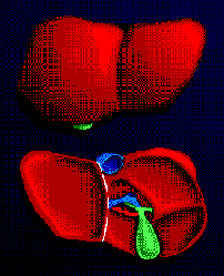
| NORMAL LIVER |
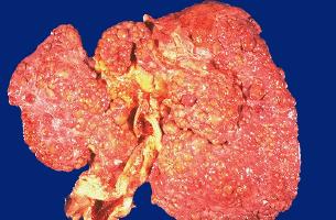
| CIRRHOTIC LIVER |