 |
(3) |
In this paper the suitability of a PTW natural diamond detector (DD) for relative and reference dosimetry of photon and electron beams, with dose per pulse between 0.068 mGy and 0.472 mGy, was studied and the results were compared with those obtained by a stereotactic silicon detector (SFD). The results show that, in the range of the examined dose per pulse the DD sensitivity changes up to 1.8% while the SFD sensitivity changes up to 4.5%. The fitting parameter, D, used to correct the dose per pulse dependence of solid state detectors, was D = 0.993±0.002 and D = 1.025±0.002 for the diamond detector and for the silicon diode respectively. The D values resulted independent of particle type of two conventional beams (a 10 MV x-ray beam and a 21 MeV electron beam). So if D is determined for a radiotherapy beam, it can be used to correct relative dosimetry for other conventional radiotherapy beams. Moreover the diamond detector shows a calibration factor independent of beam quality and particle type, so an empirical dosimetric formalism is proposed here to obtain the reference dosimetry. This formalism is based on a dose-to-water calibration factor and on an empirical coefficient, that takes into account the reading-dependence on the dose per pulse. This procedure is applicable also to silicon diode determining its calibration factor for each energy or quality of the beam.
The PTW diamond detector (type 60003)
has been widely used for relative dosimetry of photon and electron beams 1-4.
The quasi water-equivalence in terms of atomic number and the small dimensions
of the sensitive volume make the diamond detector attractive for dose measurements
involving small radiation fields that are affected by the presence of lateral
electron disequilibrium 4 and steep dose gradients. Moreover the
literature reports that the PTW diamond detector sensitivity is independent
of the photon beam quality in the range between 4 and 25 MV 5, and
of the electron beam energy in the range between 5 and 20 MeV 6.
Although this is to be expected, to be due to the near water equivalence of
diamond, an energy dependence of the PTW detector sensitivity might be introduced
by the presence of the contact materials 7.
The use of the diamond detector in reference
dosimetry has some interest because of its high resistance to radiation damage7,
whereas the silicon detector has some problems caused by radiation damage8.
Recently Mobit et al 6,
by using a Monte Carlo simulation of electron beams, have studied the applicability
of the Spencer-Attix cavity equation to determine the absorbed dose in the phantom
from the absorbed dose in the sensitive volume of the diamond detector. Their
results showed that the PTW diamond detector may be calibrated against an ionization
chamber, at the maximum dose depth, dmax, for a given electron beam
energy. This calibration factor can be used for the dosimetry of electron beams
in the energy range between 5 and 20 MeV with uncertainty of 1 %, for depths
of irradiation close to dmax, and 3% for all the other
depths. However it is known that the diamond detector has an under-response
with increasing dose rate 1,2, due to a very short electron-hole
recombination time, which decreases as dose rate increases. Fowler 9
has introduced a fitting parameter, D ,
to correct the dose rate
dependence of solid state detector reading. Heydarian et al. 2 showed that D is energy-independent
for photon beams in the range between 4 and 23 MV with an average dose rate
ranging between 0.69 Gy min-1 and 7.85 Gy min-1.
In this paper the D values
obtained for a PTW diamond detector, by using a 10 MV photon beam and two 21
MeV electron beams, are showed and compared with the D values
evaluated for a stereotactic silicon diode (the characteristics of the two solid
state detectors are described in materials and methods section). Moreover, the
sensitivity dependence on the dose per pulse has been determined for the two
solid state detectors, and for the PTW detector an empirical dosimetric formalism
for the dose to water determination is proposed here.
2.1 PTW and SFD solid state detectors
The PTW diamond detector (DD) (type 60003,
serial number 7-031) has been well documented by Rustigi et al 3. The used DD consists of a natural diamond crystal
with a sensitive area of 3.8 mm2 and 0.26 mm thick. The sensitive volume of the diamond is located
1 mm below the detector front surface of 7.3 mm in diameter. A bias voltage
of 100 V was applied and , as suggested by the detector certificate, to stabilize
the DD reading a preirradiation dose of 5 Gy was applied each time the detector
was used to obtain a set of measurements , while no preirradiation was carried
out by the manufacturer.
The stereotactic field detector (SFD) manufactured
by Scanditronix is a high-doped p-type silicon diode 10 (model DEB050
serial number 1029). The SFD sensitive area of 1.1 mm2
, 0.06 mm thick, is located 0.7 mm below the detector front surface of 4.5 mm
in diameter. The SFD was preirradiated by the manufacturer to 8 kGy and no preirradiation
is required before the experimental use.
The DD and SFD were connected to a PTW Multidos
electrometer model QC6.
2.2 Plane parallel ionization chamber
A PTW parallel plate Markus ionization chamber,
model 23343, was used as a reference dosimeter to determine the dose per pulse
of the radiation beams. The Markus ionization chamber presents: a nominal sensitive
volume equal to 0.055 cm3, a nominal electrode distance equal to
2 mm, a polarity effect less than 0.5 % and a recommended bias polarizing voltage
equal to 300 V. The Markus calibration factor, ND,P= 5.030 108
GyC-1 (±0.5%, 1s), in terms of absorbed dose to the
air cavity, was determined in a water phantom by comparison with a cylindrical
reference ionization chamber ENEA, model ESC/87 (calibrated in terms of air
kerma), applying the Italian Protocol 11 (that follows the recommendations
of the IAEA 12). The dose to water was determined by using the Italian
Protocol 11, in particular the Markus reading was corrected
by using the saturation factor (that ranged between
0.2 % and 0.6 %). The Markus ionization chamber was connected to a Keithey
electrometer model 35617.
2.3 Radiation beams and experimental set-up
The measurements were performed with a 10
MV x-ray beam with a monitor unit rate (MUR) equal to 200 MU min-1
and two electron beams of 21 MeV with MUR equal to 200 MU min-1 and
400 MU min-1. All the beams were supplied by a linac Saturne 43 G.E.,
operating in the Radiotherapy Department of the UCSC. Table I reports the MUR,
the pulse repetition frequency (PRF) and the range of the dose per pulse, for
the photon and electron beams here used. The pulse duration of the beams was
5 ms and the different dose per pulse values were obtained
by changing the source surface-phantom distance (SSD).
The aim of the photon and the electron beam
selection was to verify, for the two detectors, the independence of the D values
as well as the diamond sensitivity on the particle type. The two frequencies
supplied by the Saturne 43, were used to verify the independence of the results
on the PRF used by conventional linacs.
The measurements were carried out in a Nucletron
automatic water phantom of 60x65x67 cm3 where the three detectors
were irradiated simultaneously with a beam entering the surface of the water
phantom perpendicularly. The Markus ionization chamber was positioned on the
beam central axis, with its reference point (inner surface of the entrance plate)
placed at dmax (the depth of maximum dose) for the electron beams
and at dmax and 5 cm for the photon beam. The DD and the SFD were
positioned laterally to the ionization chamber, on right and on the left side
respectively, at the same depth in water. In this way the centers of the DD
and SFD sensitive volumes were at a distance of 3 cm from the beams central
axis; in that region the dose homogeneity was better than 0.2 % at all chosen
SSDs .
For the electron beam measurements the used
SSDs were 90 cm, 120 cm, 150 cm, 180 cm and 204 cm, for all chosen SSDs the
field size was 27x27 cm2 at dmax=3.8 cm. The energy at
phantom surface, E0, resulted equal to 19.5±0.2 MeV for all chosen SSDs.
For the photon beam measurements the different
dose per pulse values were obtained using the SSDs of 80 cm, 100 cm, 120 cm,
150 cm, 180 cm and 204 cm, and for all chosen SSDs the field size was 30x30
cm2 at the detectors depths of 5 cm and dmax=2.5 cm respectively.
As reported in section 2.2 the measurements
obtained by the Markus ionization chamber were converted in dose to water per
pulse values, Dw, (table I), taking into account the PRF values and
the irradiation time. The detectors were irradiated with a number of monitor
units (MU) sufficient to obtain an accuracy of 0.5 % on the interval time measured
by the linac clock. Dw was selected instead of the average dose to
water to compare the detector sensitivity obtained with different PRF and dose
per pulse of the used beams.
For homogeneity in this paper the Dw has been used also for the D evaluation.
3.
Results
In order to determine the relationship between
the reading per pulse of the solid state detectors, M, and the dose per pulse,
Dw, the empirical expression was used 4:
| M = aDwD | (1) |
where a is a constant and D is the
fitting parameter to correct the dose rate dependence of the detector reading
(equal to 1 for a detector with linear reading per pulse). The D values
were determined separately for each PRF and type of the particles as the
slope of the linear fit of the log normalized M against log normalized Dw.
The normalization is obtained at the values of lowest Dw1.
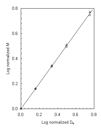
Figure 1 shows a logarithmic plot of normalized reading per pulse for DD and
SFD detectors as a function of the log normalized Dw obtained for
a 21 MeV electron beam with a PRF of 200 Hz.
The photon beam measurements were performed at dmax
and at 5 cm to evaluate the D dependence on the electron contamination
from the collimator system. The D values
turn out to be the same within the experimental uncertainties in the two experimental
conditions for both the detectors.
Table II reports the D values
of the two solid state detectors obtained by electron and photon beams. The
reading precision or reproducibility equal to 0.5% (1s) was evaluated
for the two solid state detectors while for the Markus ionization chamber resulted
equal to 0.3% (1s). The D variations
reported in table II were obtained with the method of least squares and assumed
as experimental uncertainties. Table II shows that the D values,
for each detector, are the same within the experimental uncertainties for the
two types of the particles and the PRF values examined. The D independence
of PRF was expected because, in the described experimental conditions, the charges
produced by each pulse are collected independently. Indeed the transit time,
tr, required for an electron to pass through the solid from one electrode
to the other is tr=L2/Vm, where L is the thickness of the sensitive volume, V is the operating bias
and m is the electronic mobility 9. Since the transit
times are tr = 3.8 ns for the DD and tr= 55.4 ms for the
SFD, and the smallest interval time between two pulses is about 5 ms, the charges
produced by a pulse (that are not recombined with holes) are entirely collected
before the start of the successive pulse.
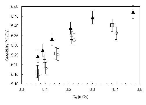
Figure 2 reports the DD sensitivity as a
function of the dose to water per pulse Dw for the two electron beams
with PRF equal to 100 and 200 Hz, and for the photon beam with PRF=100 Hz. In
this paper the detector sensitivity is defined as the ratio between the solid
state detector reading per pulse, M, and the dose per pulse, Dw,
determined by Markus chamber. The bars, 0.6 %, are obtained as the square root
combination of diamond and Markus chamber precisions (1s). Figure
2 shows that the DD has an under-response with increasing dose to water per
pulse 1,2,4 and this trend is independent of the particle type and
the PRF.
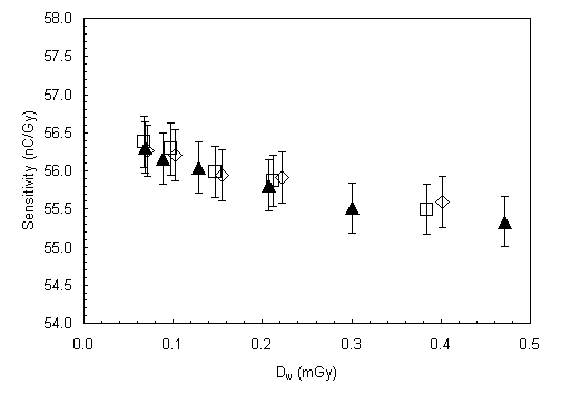
Figure 3 reports the SFD sensitivity values
as a function of the Dw values, obtained using the two electron beams
with PRF equal to 200 and 100 Hz, and the photon beam with PRF = 100 Hz. Figure
3 shows that the silicon diode has an over-response with increasing dose to
water per pulse1,2 and a sensitivity dependence on the beam quality.
This is essentially due to the non water equivalence of the silicon material
10.
Mobit et
al.6, for electron beams, and Laub et al.5, for photon beams, showed that the DD sensitivity
is energy independent, and the results reported in figure 2 show that, within
the experimental uncertainties, the DD sensitivity as a function of the dose
per pulse is also independent of the particle type. So the DD can be used for
dose measurements, only correcting the M reading per pulse for the dose per
pulse dependence. As for the non linearity of M versus Dw (equation
1) in the range from 0.068 mGy to 0.472 mGy, an empirical coefficient of linearity-rate,
Plr, was determined as the DD calibration factor (the reciprocal
of the sensitivity) Nw,D=Dw /M normalized at Nw,Dref =Dw,ref/Mref with a reference reading per pulse Mref=0.0167
nC obtained for a reference dose per pulse Dw,ref=0.300 mGy:
| Plr=[Dw /M]/ Nw, Dref | (2) |
By equation (1) Plr, may be also expressed
by the equation
|
(3) |
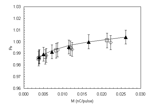
Figure
4 reports the Plr values as a function of the M values and the solid
line was obtained by the expression (3), with a value of D=0.993 that is the average value of the data reported in table II.
The DD and the SFD detectors present a reading per pulse, M, dependent on the dose per pulse, but they can be suitable to obtain the percentage depth dose, PDD(d), at depth, d, by the expression proposed by Laub et al.
| PDD(d)=[M(d)/M(dmax)]1/D 100 | (4) |
were dmax is the depth of maximum dose. The
results reported in table II show that for the examined DD and SFD detectors
the D values are, within the experimental uncertainties, independent
of the particle type, the beam quality and the PRF. However, it is important to observe that the D value has
to be determined for each PTW diamond detector or Scanditronix SFD, because
the D value is detector dependent as reported in literature7-8.
The results
reported in figure 2 show that the DD sensitivity, in a range of dose per pulse
from 0.068 mGy to 0.472 mGy (that covers the conventional radiotherapy dose
rate, measured at reference depths in water phantom for beam calibration), is
independent of the particle type and beam quality. So if Nw,Dref
and Plr are determined for a DD detector with a radiotherapy beam,
the dose per pulse at depth d, Dw(d), can be obtained, also for other
radiotherapy beams using the expression :
| Dw (d)= M(d) Nw,Dref Plr(M) | (5) |
where M(d) is the reading per pulse.
The dose to water in terms of Gy/MU may be
obtained by expression (5) multiplying Dw(d) by the number of pulses
per MU. This number is determined multiplying the PRF by the irradiation time
per MU used. So the DD may be used for reference dosimetry in those cases of
small field size and steep dose gradients, for which a ionization chamber may
be difficult to use.
The results reported in figure 3 show that the dosimetric formalism, above described, is applicable to the silicon diode examined only if the calibration factor, Nw,Dref, is determined for each quality and energy of the beam (in fact its sensitivity depends on the energy and the quality of the beam).
Acknowledgements
This work has been developed by a grant from MURST 1998
(Ministero dellUniversità e della Ricerca Scientifica e Tecnologica). We gratefully
acknowledge D. Di Nucci, P. Di Nicola, A. Porcelli for their technical assistance.
References
1 P.W. Hoban, M. Heydarian, W.A.
Beckham, and A.H. Beddoe, Dose rate dependence of a PTW diamond detector in
the dosimentry of a 6 MV photon beam, Phys. Med. Biol. 39, 1219-1229 (1994).
2 M. Heydarian, M. Zahmatkesh, P.W.
Hoban, and A.H. Beddoe, Dose rate correction factors for diamond detectors
for megavoltage photon beams, Phys. Med. 13, 55-60 (1997).
3 S.N. Rustgi, D.M.D. Frye, Dosimetric
characterisation of radiosurgical beams with a diamond detector, Med. Phys.
22, 2117-2121 (1995).
4 A. Piermattei, L. Azario, A. Fidanzio, G. Arcovito, Quasi water-equivalent
detectors for photon beams that present lateral electron disequilibrium, Phys.
Med. 14, 9-17 (1998).
5 W.U. Laub, T.W. Kaulich, and F.
Nusslin, Energy and dose rate dependence of a diamond detector in the dosimetry
of 4-25 MV photon beams, Med. Phys. 24, 535-536 (1997).
6 P.N. Mobit, G.A. Sandison, An
EGS4 Monte Carlo examination of the response of a PTW-diamond radiation detector
in megavoltage electron beams, Med. Phys. 26, 839-844 (1999) .
7 B. Planskoy, Evaluation of diamond radiation dosimeter, Phys. Med. Biol.
25, 519-532 (1980).
8 D. Wilkins, X.A. Li, J. Cygler,
and L. Gerig. The effect of dose rate dependence of p-type silicon detectors
on linac relative dosimetry. Med. Phys. 24 , 879-881 (1997) .
9 J.F. Fowler, Solid state electrical
conductivity dosimeters Radiation Dosimetry ed F.H. Attix and W.C. Roesch,
(New York : Academic) (1966).
10 G. Rikner, Silicon diodes as detectors in relative dosimetry of Photon,
electron and proton radiation fields, (Sweden : Uppsala Universitet) (1983).
11 Associazione Italiana Fisica Biomedica (AIFB), Protocollo
per la dosimetria di base nella radioterapia con fasci di fotoni ed elettroni
con Emax fra 1 e 40 MeV, Notiziario della A.I.F.B. (VI), 2 (1988)
12 International Atomic Energy Agency (IAEA), Adsorbed dose determination
in photon and electron beams: An international code of practice, Technical Reports Series No. 277,
Vienna, (1987).
TABLES
Table I . Monitor unit rate (MUR), pulse repetition frequency (PRF) and range
of dose per pulse, for photon and electron beams, supplied by a linac Saturne
43 G.E.
Radiation
beam
|
MUR (MU min-1) |
PRF (Hz) |
Range of dose
per pulse (mGy) |
|
Photon 10 MV
|
200
|
100
|
0.069-0.472
|
|
Electron 21 MeV
|
200
|
100
|
0.071-0.400
|
|
Electron 21 MeV
|
400
|
200
|
0.068-0.384
|
oooooTable II . Dose rate correction factor, D, values obtained for the diamond detector (DD) and the stereotactic field detector (SFD) by photon and electron beams.
|
Detector
|
Photon (100 Hz) |
Electron
|
Electron
|
|
DD
|
0.994±0.002
|
0.994±0.002
|
0.991±0.002
|
|
SFD
|
1.023±0.002
|
1.025±0.002
|
1.026±0.002
|
| Figure 1. Log normalized values of reading per pulse,
M, for diamond detector (DD) (¡) and steretactic field detector (SFD) (r), as a function of the log normalized values of dose
per pulse, Dw, obtained for a 21 MeV electron beam with pulse
repetition frequency of 200 Hz. The normalization is obtained at the lowest
values of Dw and M. |
| Figure 2. Diamond detector sensitivity
as a function of dose per pulse, Dw, obtained for the 21 MeV
electron beams with pulse repetition frequency (PRF) of 200 Hz (£) and 100 Hz (¯), and for the 10 MV photon
beam with PRF=100 Hz(p). |
| Figure 3. Stereotactic field detector sensitivity as
a function of dose per pulse, Dw, obtained for
the 21 MeV
electron beams with pulse repetition frequency (PRF)
of 200 Hz (£) and 100 Hz (¯), and for the 10 MV photon beam with PRF=100 Hz(p). |
| Figure 4. Plr values for diamond detector obtained with 21 MeV electron beams with pulse repetition frequency (PRF) of 200 Hz (£) and 100 Hz (¯) and with a 10 MV photon beam with PRF=100 Hz (p). The Plr values are reported as a function of reading per pulse, M, and normalized at Mref=0.0167 nC. The solid line, Plr (M,D), was obtained by expression (3) with an average D value of 0.993. |
 |
 |
||
|
Figure
1
|
Figure
3
|
||
 |
 |
||
|
Figure
2
|
Figure
4
|
oooo
oooo