Somebody circled in grease pencil the "ovoid of enhancement". ( Also called a "hot spot". )
The original tumor was biopsied to be a grade 3 (malignant) so this area of enhancement was considered "worrisome for a recurrent tumor". Meaning oncologists felt it was likely the tumor growing back from residual cancer cells that were not removed during the surgery.
One surgeon did point out that it might be harmless "post surgical inflammation". That little spot caused a lot of anxiety, but Praise God, it didn't amount to anything.
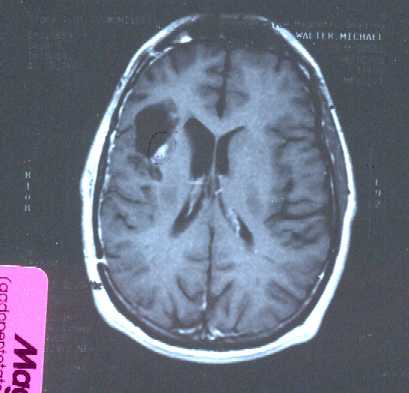
Same day.
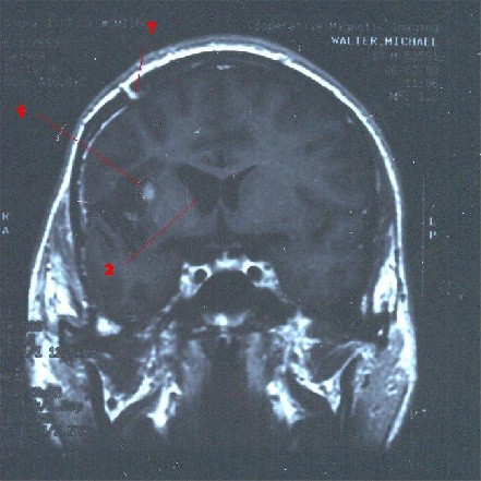
Different Angle.
I tried to add some labels to this picture which are not very clear.
The most upper one is a ?
What it is pointing to I do not exactly know. But this is the general area where the bone flap was that provided access to my brain for the tumor resection.
The label in the middle is a 1.
Pointing to the "hot spot".
The lower label is a 2.
pointing to these pockets that everyone has in their brain which I forget their name. I believe they function as reserviors for the cerebro-spinal fluid which flows around the brain. What is interesting is that the one on the tumor side of the brain is larger. This is probably becase the brain is shifting around as it slowly fills in the void left from the tumor..
Table of Contents.