This is only 4 of the original 158 pictures that were taken on Nov. 19, 2002. You can see my eyes in the pics on the right. Therefore you know that the front of my head is toward the left in all pics.
The tumor was postioned in the RIGHT FRONT of my brain, although it would appear to be on the left side of my brain from these pics. I think that is because us lay people generally think of looking at pictures, drawings and maps from a "bird's eye view".
MRIs are not necessarily presented that way . . . so they can be confusing to read.
The MRI usually has coding by the image to give information for interpretation and orientation (that is which way is right, which way is left?)
That coding is too faint and small to show up in the scan below, so I have included labels for orientation showing:
R Right
L Left
A "Anterior" which means Front
P "Posterior" which means Rear
I know the tumor was on the right side because they told me it was and because that is the side of my head that was operated on.
Also my symptoms were on the left side of my body. During my seizures, which were minor, I felt numbess in my left hand and left side of my face. And as we remember from Biology class the Right side of the brain controls the Left side of the body and vice versa.
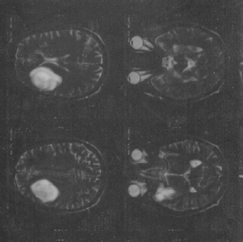
The tumor measured 6.1 x 4.4 x 4.3 cm.
1 inch equals 2.54 cm so in inches the tumor was about 2.4" x 1.7".
So it was slightly smaller than a tennis ball which has a diameter of about 2.5 " .
Table of Contents.