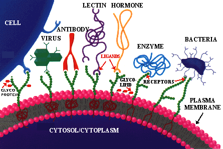
The universal principle of molecular recognition. This should be very familiar to you by now, so it may turn up on exams. Every cell contains membrane-bound molecules exposed to the environment which communicate with it. These molecules act as the "eyes, ears and nose" of a cell. That is, they "see", or "smell" what other molecules are out there like a hunting dog winds its prey. They contain, as part of each molecule, specific regions on each molecule called RECEPTORS or BINDING SITES. Molecules in the environment contain specific components called LIGANDS that have the characteristic of binding or attaching (docking with) specifically at unique sites on the cell's receptor molecules. Following this attachment a number of different responses may occur. In some cases a message is passed to the interior of each cell as to the situation it has found, while in others the attached molecule may be transported into the cell. In the former case this information, in turn, triggers the cell to carry out a series of preprogrammed responses based on the data it has received; this is analogous to the Mars rover receiving commands from earth. We will discuss some of these responses throughout the course. Permission to use this cartoon was granted by Sigma Chemical Co.
![]()
USING PATHOGENIC BACTERIA TO FIGHT CANCER
GENE THERAPY TO FIGHT CYSTIC FIBROSIS
GENE THERAPY (SMART BOMB) TO FIGHT CANCER
![]()
As previously discussed, we are in the middle of a biological informational revolution that will change our lives in ways we can only begin to imagine. Although Molecular biology and Genetic Engineering are perplexing to most people (e.g. the jurors on the O.J. trial) their basic principles are straightforward enough so that any person who would consider themselves to be "educated" should understand them. The basic laboratory steps in GENETIC ENGINEERING (go to "Educational Leaflets") are so simple that high school students routinely perform them. In this section of Micro 101 you will become acquainted with these principles, the fundamental steps of genetic engineering and some of the things that can be done using these procedures. The objectives of this section are to learn:
![]() What the BASIC TOOLS
of genetic engineering are and how they work.
What the BASIC TOOLS
of genetic engineering are and how they work.
![]() How GENE CLONING
is carried out.
How GENE CLONING
is carried out.
![]() How DNA can be used to IDENTIFY all
living organisms
How DNA can be used to IDENTIFY all
living organisms
![]() How we can DETECT
very TINY QUANTITIES
of DNA even when they may be millions of years old.
How we can DETECT
very TINY QUANTITIES
of DNA even when they may be millions of years old.
| On 7/11/97 it was reported that analysis of DNA from a Neanderthal's skeleton showed that they were a different species from us. So if you're ever told you're acting "like a Neanderthal" you can tell them "no I'm acting like a typical Homo Sapiens". |
![]() In addition, ETHICAL
problems of the genetic engineering revolution will be presented and some
possible ways of dealing with them considered.
In addition, ETHICAL
problems of the genetic engineering revolution will be presented and some
possible ways of dealing with them considered.
![]()
MOLECULAR BIOLOGY is the area of biology that is concerned with how biological molecules #INTERACT with each other at the MOLECULAR LEVEL; a sort of ONE-ON-ONE SITUATION. To fully understand the PROCESSES OF LIFE it is necessary to understand what is occurring at the atomic/molecular level. Life is a matter of TINY, but specific, interactions occurring at ATOMIC DISTANCES. Since we are currently unable to observe directly the molecular events taking place on an enzyme's or membrane's surface, or between a ribosome and mRNA etc., we must investigate these processes through indirect means. This is analogous to putting you in a dark room with a number of machines and asking you to figure out how they work together. However, there are scientific procedures in development that will eventually allow us to observe molecular processes while they are occurring within a cell (I hope I live long enough to "see" that!). A molecular biologist, which almost EVERYONE in biological science today calls themselves, uses a variety of SENSITIVE procedures to probe (examine) molecular interactions. These procedures involve using LABELS or REPORTERS to follow molecules through the processes they undergo and to pinpoint their presence, location and amount within a cell. There are MACHINES, some of which are only a cm square, that can detect and quantitate INDIVIDUAL MOLECULES of protein, DNA or RNA within a few minutes. There are procedures that allow scientists to MAGNIFY, or amplify very small quantities of biological materials so that they can identify and study them. Techniques are currently available that allow us to CONTROL A CHANGE anywhere in the genetic code of a gene. There are microscopes that allow us to see individual atoms and even groups of electrons. These machines, techniques and reagents are expensive, the experimental procedures labor intensive and, because of the EXQUISITE SENSITIVITY of the techniques, prone to error. However, their prices are dropping and they are becoming more reliable.
I have listed some of the changes you are likely to experience within your lifetime, indeed some within the next few years. With the sequencing of the ENTIRE HUMAN GENOME anticipated within the next 3 years (as announced by the federal organization funding the project in Oct 1998), a whole new set of possibilities will be unlocked. Currently (Fall 1998) we have sequenced ~3% of the human genome, and the rate of sequencing is accelerating as sequencing techniques are improved. About 3,000 human genetic diseases are currently known. This means that approximately 14% of newborns are afflicted with a "visible" genetic disease. This does not begin to include the genetic-based "tendencies" (to develop cancer, arthritis, TB, colds, asthma, flu etc.) and/or "conditions" which all of us have; what genetic conditions do you have (e.g. shy, migraines, depression, fair skin)? For example, I and my half brother suffer from a genetic condition called Celiac Sprue, which means that wheat protein (gluten) does bad things to the cells in our intestine and we can't eat pizza etc. However, we are FINE as long as we don't eat gluten-containing products. Sprue is a common genetic "condition" that effects up to 25% of the Irish and lesser numbers of other ethnic groups. How many reading this are Irish? Some of you suffer allergies, nearsightedness, migraines, etc., all of which have their basis in your genome. Once the human genome is mapped we can not only identify the particular alleles each of us have, but we will eventually figure a way to replace "undesirable genes" with "good genes".
One current problem with the genetic revolution is that knowing which gene causes a disease condition and being able to identify the presence of that gene in a person, doesn't mean we UNDERSTAND the molecular biology of the disease process, thus we usually CAN'T PREVENT or yet CURE a disease caused by a defective gene. An example of this TERRIBLE DILEMMA is that a number of genes that predispose women to breast cancer have been discovered (and new ones are continually being found). This raises important ethical and personal considerations. It is estimated that each human carries an average of three lethal genes (genes which become lethal in an offspring who receives two copies of the gene).
![]() If a CURE or PREVENTION is NOT POSSIBLE,
should a person be told they carry such a gene?
If a CURE or PREVENTION is NOT POSSIBLE,
should a person be told they carry such a gene?
![]() If a cure or prevention is NOT POSSIBLE,
would you want to know you had a time-bomb ticking in you?
If a cure or prevention is NOT POSSIBLE,
would you want to know you had a time-bomb ticking in you?
![]() If a cure or prevention of a genetic predisposition to breast cancer is NOT
POSSIBLE, would you, as a woman, consider having
your BREASTS REMOVED to prevent getting the disease? If you decide to do this
(and many women have), without any sign of cancer, should the Health Insurance
Co. pay for the removal of a healthy breast because it is "elective"
surgery?
If a cure or prevention of a genetic predisposition to breast cancer is NOT
POSSIBLE, would you, as a woman, consider having
your BREASTS REMOVED to prevent getting the disease? If you decide to do this
(and many women have), without any sign of cancer, should the Health Insurance
Co. pay for the removal of a healthy breast because it is "elective"
surgery?
![]() What if it were testicular cancer? What would you males do?
What if it were testicular cancer? What would you males do?
| Let's examine one more "gray-zone" situation before proceeding. Manic depression (MD) is an illness that strikes many people. In some it is relatively mild while in others it takes a terrible toll, often leading to SUICIDE. It is also appears to have a genetic basis as it runs in families, and often stigmatizes affected and unaffected members alike. I would wager money that most of you reading this know someone who is a manic depressive and, of course some of you are manic depressives. Yet, the disease is often treatable by inexpensive drugs and psychotherapy, although many people suffer torment for years before they are properly diagnosed and treated. An interesting side of manic depression is that many of the creative artists and politicians have been (maybe some of our current politicians are manic depressive) manic depression. Robert Schumann, Lord Byron, Vincent van Gogh, Winston Churchill and Alfred, Lord Tennyson all (apparently) suffered classical symptoms of manic depression (see "Manic-depressive illness and creativity", Sci. Am. Feb. 1995 & Nov. 1995). Once we identify the genes causing manic depression, we can use this information to make decisions about whether to bare children carrying this gene or to ABORT any fetus carrying this gene. What would you do? Eventually we will be able to completely cure (repress) the condition. Will the ramification of these findings make for a less interesting and stimulating world? What is your opinion? |
Another weighty issue is: As we become able to screen for the presence of even more DEFECTIVE GENES, how should the information obtained be used? Consider the following questions carefully.
![]() Should one's mate, boss, insurance Co., potential employer etc. have access to
your genetic information? Can you think of situations where it would be logical
and reasonable for others to have this information? How about the army? The CIA?
Did you know that the armed forces take blood from each serviceperson for
genetic analysis?
Should one's mate, boss, insurance Co., potential employer etc. have access to
your genetic information? Can you think of situations where it would be logical
and reasonable for others to have this information? How about the army? The CIA?
Did you know that the armed forces take blood from each serviceperson for
genetic analysis?
![]() If you owned a life insurance Co. would you give your political donations to a
congressperson who favored requiring potential policy holders to give this
information to their insurance Co.
If you owned a life insurance Co. would you give your political donations to a
congressperson who favored requiring potential policy holders to give this
information to their insurance Co.
![]() Who pays for the screening for disease-genes, the cost of which currently is
high? Should I have to pay for your screening & vice versa?
Who pays for the screening for disease-genes, the cost of which currently is
high? Should I have to pay for your screening & vice versa?
![]() What if you control the genetic information and you know that giving it out will
lead to an abortion; what would you do?
What if you control the genetic information and you know that giving it out will
lead to an abortion; what would you do?
![]() What if you have access to the genetic information of someone your daughter/son,
sister/brother etc. is going to marry and you don't think they've told them of a
serious genetic "condition". Would
you tell? Who owns your loyalty? What
is ethical here?
What if you have access to the genetic information of someone your daughter/son,
sister/brother etc. is going to marry and you don't think they've told them of a
serious genetic "condition". Would
you tell? Who owns your loyalty? What
is ethical here?
A final reminder of anyone who is considering avoiding these problems; gene therapy trials are currently underway (but they're having problems working: TIME 10/9/95; Science 269:1050 [1995]), people are deciding to abort on the basis of the results of fetal genetic testing and the number of identified defective genes increases almost daily (and some of the ones they find will be ones you and I carry).
Click on the URLs below for perspectives on genetic engineering ethics:
![]()
By the early 1960s geneticists had a good basic picture of genetics. However, they did not have any way to obtain and study the genes themselves except through the laborious procedures of mating and testing mutants. These procedures take an immense amount of time, money and effort and yield disproportionately MEAGER RESULTS. Further, there were no techniques to examine the genes at a molecular level, yet unless this was done the ultimate questions could never be answered. Because of these problems older geneticists were leaving the field and younger, ambitious ones, were looking elsewhere for their creative outlet. Then, as so often happens in science, a series of TOTALLY UNRELATED SERENDIPITOUS DISCOVERIES were made which propelled the genetic revolution upon the world. It all started with a seemingly odd result obtained with some phage.
In the 1950s it had been noted that if one grew a particular #bacteriophage on a particular bacterial mutant host strain (A) and then infected a bacterial mutant-strain (B), of the same species, with the phage A, the yield of phage from strain B was VERY LOW. However, if you took the few phage B that were produced and infected strain B with them, the phage yield was now NORMAL. Subsequently, in the 1960s, it was found that the phage DNA that grew on strain B had been CHEMICALLY MODIFIED so that it could not be cleaved (destroyed) by a DNase in strain B. The DNase involved in this cleavage was found to be somewhat specific, in that it mainly cut the DNA at CERTAIN SEQUENCES; unless these sequences had been CHEMICALLY MODIFIED by enzymes in the cell (missing in strain A). All previous DNases cut DNA randomly, so this finding suggested that DNA could be cut up into SPECIFIC FRAGMENTS which would ALWAYS contain the SAME SET OF GENES between the cut-sites. However, these first "specific" DNases did not prove as SPECIFIC as hoped. Finally, in 1970 Hamilton Smith accidentally found that a DNase from the bacterium, Haemophilus influenzae, CUT DNA ONLY at UNIQUE DNA SEQUENCES known as PALINDROME SITES.
![]()
The term palindrome refers to groups of letters that form the same words when read both forward and backward. For example, "MADAM, I'M ADAM"; "ABLE WAS I ERE I SAW ELBA"; "DENNIS SINNED"; "MA IS A NUN, AS I AM"; "SEX AT NOON TAXES".
![]()
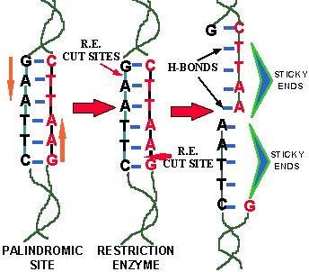
In DNA a PALINDROME SITE is a SEQUENCE OF BASE PAIRS in double stranded DNA that reads the same backwards and forward across the double strand. For example, the sequence of base pairs GAATTC is a palindrome because both sequences of the double strand READ THE SAME when read from either their respective "G" or "C" ends (COMPLEMENTARY strand = CTTAAG). The enzymes that cut these specific sites are called RESTRICTION ENZYMES. Based on the TYPES OF CUTS they make, there are two types of restriction enzyme:
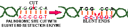
![]() One group cuts straight across the double stranded
DNA, producing BLUNT ENDED DNA
(Fig.
2).
One group cuts straight across the double stranded
DNA, producing BLUNT ENDED DNA
(Fig.
2).
![]() Another type cuts the strand of DNA
OFF THE CENTER of the palindrome sites, but between
the SAME TWO BASES
on the opposite strands. This leaves one or more bases overhanging on each
strand and such ends are called STICKY
ENDS (Fig. 1) because they form HYDROGEN
BONDS with their complementary cut counterparts.
Another type cuts the strand of DNA
OFF THE CENTER of the palindrome sites, but between
the SAME TWO BASES
on the opposite strands. This leaves one or more bases overhanging on each
strand and such ends are called STICKY
ENDS (Fig. 1) because they form HYDROGEN
BONDS with their complementary cut counterparts.
For example, in the base sequence GAATTC, the restriction enzyme that cuts this site cleaves the DNA between the "G" and the "A" on each complementary strand, thus leaving the overhangs of AATT & TTAA respectively (Fig. 1). Over 200 different restriction enzyme have been isolated that cut at many different specific DNA sequences. They are all QUITE SPECIFIC and this specificity is the KEY to their use. Restriction enzymes that cut at 4, 5, 6, 8, 9, and 11 base pair palindrome sites have been found. It follows that the MORE BASE PAIRS in the palindrome site the LESS LIKELY a site is to exist statistically. That is, a 4-base-cutting restriction enzyme cuts the same DNA many more times than an 8-base-cutter does. The odd numbered cutting sites are not true DNA palindromes, in that the central base pair reads different in the two directions. For example AA?TT is a 5 base pair cutting site, where the ? = a number of different bases.
|
|
![]()
![]() CRITICAL THINKING QUESTION: The following are examples of restriction enzyme DNA
sites on a single strand. Add the complementary bases and find the various
restriction enzyme sites within each sequence: CCTAGT; AATCCTAGGACG; AAATTAATCGG;
TAAGGCGCGCCTAAT;
CRITICAL THINKING QUESTION: The following are examples of restriction enzyme DNA
sites on a single strand. Add the complementary bases and find the various
restriction enzyme sites within each sequence: CCTAGT; AATCCTAGGACG; AAATTAATCGG;
TAAGGCGCGCCTAAT;
![]()
![]()
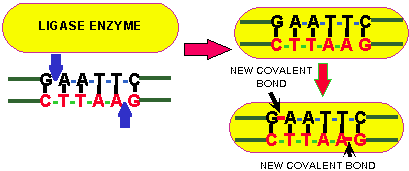
The final important component in genetic engineering is an enzyme called LIGASE. Ligase is an enzyme that covalently joins the sugar-phosphate backbone of bases together. Ligase is also involved in DNA replication. In effect LIGASE reverses the action of restriction enzymes, which break the sugar-phosphate backbone-bonds. Ligase requires energy to form these bonds (Fig. 3). Ligase will join either "sticky" ends or "blunt" ends, but it is more efficient at closing sticky ends because the "sticky overhangs" of these ends adds stability which holds the strands together while the ligase works, whereas the blunt ends are LESS STABLE. The sticky ends must have the SAME "base overhang" (Fig. 1), but ANY BLUNT END can be joined to any other blunt end (Fig. 2).
Review of the components of genetic engineering:
![]() (1)
Double-stranded DNA contain palindrome restriction enzyme sites.
(1)
Double-stranded DNA contain palindrome restriction enzyme sites.
![]() (2)
Many types of restriction enzymes cut or cleave palindrome sites in the
double-stranded DNA sample forming either a sticky or a blunt end.
(2)
Many types of restriction enzymes cut or cleave palindrome sites in the
double-stranded DNA sample forming either a sticky or a blunt end.
![]() (3)
The enzyme ligase plus an energy source fuses or joins two sticky or two blunt
ends of DNA together.
(3)
The enzyme ligase plus an energy source fuses or joins two sticky or two blunt
ends of DNA together.

Animation of the ligation process.
When the sticky ends of two DNA fragments come together and hydrogen
bond through the A-T and G-C pairing, the enzyme LIGASE will form covalent bonds
between the two fragments.
![]()
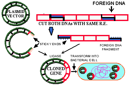
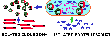
In genetic engineering the idea is to move a "GENE OF INTEREST" from its original or SOURCE DNA molecule into another or TARGET DNA molecule for some SPECIFIC PURPOSE. Those of you who are computer aficionados will note the similarity in terminology in describing the moving of computer files. We will discuss the various purposes later. The process of moving a gene from one DNA molecule to another DNA molecule is called CLONING. In the cloning process a fragment of DNA, containing a gene- or sequence-of-interest, is CLONED (moved) INTO A VECTOR where it can subsequently be grown (COPIED) in large quantities and MANIPULATED in a variety of ways. The most common vectors are bacterial PLASMIDS, but viruses and self-replicating units in eukaryotic cells are also employed as vectors. The process of cloning is straight forward and only requires the following four steps and can be done in a couple of hours or less.
![]() Isolating the SOURCE
and VECTOR
DNA. The DNA's must be relatively free of contaminating materials which
interfere with the subsequent enzymatic steps.
Isolating the SOURCE
and VECTOR
DNA. The DNA's must be relatively free of contaminating materials which
interfere with the subsequent enzymatic steps.
|
|
![]() Both the source and vector DNA are cut with RESTRICTION
ENZYMES. When sticky ends are formed the DNA is cut
with the same restriction enzyme(s), but restriction enzyme that produce blunt
ends also work well in cloning.
Both the source and vector DNA are cut with RESTRICTION
ENZYMES. When sticky ends are formed the DNA is cut
with the same restriction enzyme(s), but restriction enzyme that produce blunt
ends also work well in cloning.
![]() The vector and source DNA are mixed with a LIGASE
SYSTEM and covalently bonded together.
The vector and source DNA are mixed with a LIGASE
SYSTEM and covalently bonded together.
![]() Finally, the LIGATED DNA
is TRANSFORMED
into a host cell. Usually the host cell is a COMPETENT bacterium, but increasingly eukaryotic cells are being used. After suitable
growth has occurred the host cells are examined for the PRESENCE of the source
or CLONED DNA
in its cytoplasm.
Finally, the LIGATED DNA
is TRANSFORMED
into a host cell. Usually the host cell is a COMPETENT bacterium, but increasingly eukaryotic cells are being used. After suitable
growth has occurred the host cells are examined for the PRESENCE of the source
or CLONED DNA
in its cytoplasm.
The entire cloning process often takes LESS THAN A DAY and is carried out using volumes of 1,000 microliters or less. The examination of the transformed host for the gene-of-interest may take an additional day or so. A gene which has been successfully transferred in this way is said to have been CLONED. The process is referred to as "CLONING", "RECOMBINANT DNA TECHNOLOGY", or "RECOMBINANT DNA". These steps are summarized in the figure 4 below.
![]()
To do molecular biology on a single gene it is necessary to ISOLATE it from all the other genes. This is not always easy considering that an average bacterium contains 1,000 to 4,000 genes, while eukaryotic cells may contain >100,000 genes. A cloned gene can be AMPLIFIED by making many COPIES of the clone so as to produce large enough quantities to study it in detail. The usual things that are done to an amplified gene are:
![]() SEQUENCING:
Sequencing determines the base pair sequence of a gene. By reading the 3-letter
code, sequencing also describes the AMINO
ACID SEQUENCE translated from that gene.
SEQUENCING:
Sequencing determines the base pair sequence of a gene. By reading the 3-letter
code, sequencing also describes the AMINO
ACID SEQUENCE translated from that gene.
![]() MUTATION: The
gene base pair sequence can be changed in specific ways and the modified gene
can be inserted back into its original host to see what each specific mutation
does. Whereas, spontaneous mutation is random, techniques are now available that
make it possible to CHANGE
any codon within a gene to any other codon. Therefore, it is possible to study
the effects of SINGLE AMINO ACID
CHANGES on the function of the gene product, which is,
after all, the ultimate purpose of the exercise.
MUTATION: The
gene base pair sequence can be changed in specific ways and the modified gene
can be inserted back into its original host to see what each specific mutation
does. Whereas, spontaneous mutation is random, techniques are now available that
make it possible to CHANGE
any codon within a gene to any other codon. Therefore, it is possible to study
the effects of SINGLE AMINO ACID
CHANGES on the function of the gene product, which is,
after all, the ultimate purpose of the exercise.
![]() To replace a mutated form of the gene in the original host cell with a healthy
form of the gene to see exactly what it does in its intended place of residence.
To replace a mutated form of the gene in the original host cell with a healthy
form of the gene to see exactly what it does in its intended place of residence.
|
|
![]() To use the amplified gene to make HUMONGOUS
QUANTITIES of the GENE
PRODUCT for commercial purposes. This is how products
like human insulin, human growth hormone, plasminogen activator, interlukins and
the bovine milk hormone are all produced.
To use the amplified gene to make HUMONGOUS
QUANTITIES of the GENE
PRODUCT for commercial purposes. This is how products
like human insulin, human growth hormone, plasminogen activator, interlukins and
the bovine milk hormone are all produced.
![]() To INSERT the gene into ANOTHER SPECIES for some purpose. Such animals or plants
are said to be TRANSGENIC.
For example, many crops are protected against caterpillar larvae because a gene
from a bacterium has been inserted into their cells. This gene produces a
protein that is HIGHLY, and SPECIFICALLY, TOXIC to the larvae of many moths and
butterflies that attack food crops.
To INSERT the gene into ANOTHER SPECIES for some purpose. Such animals or plants
are said to be TRANSGENIC.
For example, many crops are protected against caterpillar larvae because a gene
from a bacterium has been inserted into their cells. This gene produces a
protein that is HIGHLY, and SPECIFICALLY, TOXIC to the larvae of many moths and
butterflies that attack food crops.
![]()
Transgenic means "ACROSS SPECIES" (go to "Educational Leaflets"). The above techniques give us the ability to move viral genes into humans, human genes into plants, plant genes into viruses etc. etc. There is (SPRING 96) a flood of transgenic plants in the approval pipeline; two types of transgenic tomatoes are in our local supermarkets, transgenic squash are coming soon and a number of transgenic crops are currently protected from insect larvae by the protein described above. Click here for a discussion of the potential of transgenic life; here for another cartoon and here for a site that discusses the ethics of genetic engineering.
There is a DOWN SIDE
of genetic engineering. The same procedures that can be used to produce a better
crop, or human insulin, can also be employed to make a more POWERFUL
PATHOGEN. There is evidence that some countries have
or are considering doing this. For example, Iraq was apparently growing
bacterial pathogens for ![]() biological
warfare, but it was deterred from using them when we threatened atomic
retaliation. It is only a small jump in "reasoning" to consider
engineering a "better pathogen" with which to destroy a hated
enemy (fill in blank--the news report that some Americans. consider
anyone working for the government to be the enemy). For more information read
"The spectra of biological
weapons" Scientific Am. Dec. 1996, pg. 60.
biological
warfare, but it was deterred from using them when we threatened atomic
retaliation. It is only a small jump in "reasoning" to consider
engineering a "better pathogen" with which to destroy a hated
enemy (fill in blank--the news report that some Americans. consider
anyone working for the government to be the enemy). For more information read
"The spectra of biological
weapons" Scientific Am. Dec. 1996, pg. 60.
http://hopkins.med.jhu.edu/NewsMedia/newsmedi.html Topical medical information.
http://tbase.jax.org/docs/databases.html Transgenic research data base.
http://darwin.ceh.uvic.ca/bigblue/bigblue.htm Transgenic animal data base.
http://www.cenargen.embrapa.br/binas/Library/ucs/ Union of Concerned Scientists: Problems with transgenic crops.
http://www.ext.vt.edu/news/periodicals/cses/1997-05/1997-05-03.html Transgenic cotton plants
http://ci.mond.org/9510/951010b.html Genes in milk.
http://ci.mond.org/9510/951010c.html Genes in farm animals to improve health of.
![]()
The THREE basic principles required to understand DNA fingerprinting and all that follows from it you already know (the principle of ligand/receptor binding; see the figure at the beginning of this chapter), but it is worth taking a moment to review them.
![]() BASE-PAIRING
of AT (AU) & GC is the BASIC PRINCIPLE of this procedure. If you understand
this all else fall easily into place.
BASE-PAIRING
of AT (AU) & GC is the BASIC PRINCIPLE of this procedure. If you understand
this all else fall easily into place.
![]() SPECIFICITY of enzyme activity is the second CRUCIAL principle to understanding DNA
fingerprinting. This refers to the cutting of DNA by specific RESTRICTION
ENZYMES at UNIQUE palindrome sequences.
SPECIFICITY of enzyme activity is the second CRUCIAL principle to understanding DNA
fingerprinting. This refers to the cutting of DNA by specific RESTRICTION
ENZYMES at UNIQUE palindrome sequences.
![]() The recognition that a CHANGE
in a SINGLE BASE PAIR (a mutation)
can either MAKE a restriction enzyme-site where one did not exist previously or
it can REMOVE
or ELIMINATE
a restriction enzyme site from a gene. An analogy would be to "mutate"
your phone number by one letter; callers would get a different person.
The recognition that a CHANGE
in a SINGLE BASE PAIR (a mutation)
can either MAKE a restriction enzyme-site where one did not exist previously or
it can REMOVE
or ELIMINATE
a restriction enzyme site from a gene. An analogy would be to "mutate"
your phone number by one letter; callers would get a different person.
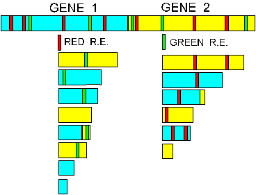
To understand DNA fingerprinting the double stranded DNA molecule must be viewed as a chain with RANDOM RESTRICTION ENZYMES-sites (palindromes) located along its length. If each of these various unique restriction sites were marked with a different color, the DNA would appear as a random multicolored stripped ribbon. If one set of unique restriction enzyme sites were colored RED and the appropriate RED-restriction enzyme added, the DNA would be CLEAVED into a series of VARIOUS SIZED FRAGMENTS depending on where the RED-SITES were located along the DNA molecule (Fig. 6). It is easy to understand that the group of DIFFERENT SIZED FRAGMENTS would be UNIQUE for two DNA strands with the RED-restriction sites located at DIFFERENT PLACES along their respective DNA double strands. The addition of a GREEN-restriction enzyme that cuts GREEN-SITES would produce a different group of sized fragments. Each of these groups of restriction enzyme-produced fragments would represent a unique FINGERPRINT of that DNA (Fig. 6).
The DNA fragments are visualized using the technique known as GEL ELECTROPHORESIS. Briefly, porous gels composed either of a PLASTIC called ACRYLAMIDE or of a derivative of agar, called AGAROSE, are used to separate DNA fragments based on size. The gels are prepared with wells into which the DNA fragments are ADDED. The gels are submerged, under an electrolyte buffer solution between a positive and a negative electrode; the DNA-containing solutions are added to the wells and the current is turned on. The DNA fragments are NEGATIVELY CHARGED so the wells containing them are placed closest to the negative electrode. When the current is turned on the DNA moves through the pores in the gel TOWARDS THE POSITIVE ELECTRODE. The shorter fragments move FASTEST because they are able to navigate through the pores of the gel more easily, whereas the longer DNA fragments move PROPORTIONALLY MORE SLOWLY through the pores. The result is a separation of the DNA fragments based on their length (SIZE). The DNA are stained by dyes that binds to the DNA and fluoresces strongly when exposed to UV-light. The various sized DNA fragments appears as a series of bands that resemble a BAR CODE. See Fig. 13.
|
|
Recently (Sci. 278:1407[1997]) the FBI has announced that DNA fingerprinting is now good enough to identify individuals with the same accuracy as classical fingerprinting because of the improvements of the procedure described above. The only problem now lies in human error; I don't think we'll ever solve that one, do you?
![]() Click here (go to "Educational
Leaflets") for a good description of DNA fingerprinting and the use of
probes.
Click here (go to "Educational
Leaflets") for a good description of DNA fingerprinting and the use of
probes.

Summary animation of the running of an agarose gel. Click here for a more detailed animation (using Chime) that lets you load the gel yourself.
![]()
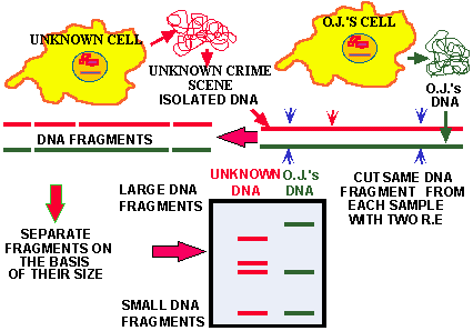
DNA fingerprinting works because of mutations which CHANGE THE GENETIC CODE, randomly MAKING and ELIMINATING restriction enzyme sites randomly throughout the DNA. Because of these variation in genes between individuals the likelihood that the restriction enzyme fragment pattern of two individuals BEING THE SAME is very very small unless they are IDENTICAL TWINS. To determine a DNA fingerprint a small amount of DNA-containing material from an organism is required. This could be obtained from any tiny quantity of tissue or blood, even the material from a single hair follicle is sufficient. The variation in the gel fragments from different DNA from members of the SAME SPECIES is called RESTRICTION FRAGMENT LENGTH POLYMORPHISM ANALYSIS (RFLP).
In Figure 7 the gel pattern of a hypothetical analysis of O.J.'s DNA vs. a sample of unknown DNA is depicted. The two isolated DNA were treated with the same restriction enzyme and the fragments separated on an agarose gel. Two unique restriction enzyme fragment patterns are seen. The conclusion is that the blood samples came from DIFFERENT INDIVIDUALS. The SOURCE of the DNA clearly influences the significance of these results. For example, if the two samples were collected at the scene of the crime it would mean one thing, but if the unknown DNA sample came from O.J.'s shirt and it matched that of one of the victims at the scene of the crime, the data takes on more significance.
|
|
![]()
FAQ: "If the genome of humans contains so many genes it must have thousands of restriction enzyme sites which would produce thousands of bands which would be very difficult to RESOLVE INTO INDIVIDUAL BANDS in a small gel wouldn't it?"
ANSWER: This is exactly what happens. When genomic DNA is treated with a restriction enzyme and separated on a gel, what is seen is a continuous smear composed of 1,000s of individual DNA fragments. This problem is solved by a technique known as HYBRIDIZATION.
Hybridization again depends on the basic principle of HYDROGEN BONDING between GC & AT bonds in nucleic acids. The principles in the hybridization steps are:
![]() DNA strands are SEPARATED
by breaking the hydrogen bonds between the bases with heat to produce SINGLE
STRANDS.
DNA strands are SEPARATED
by breaking the hydrogen bonds between the bases with heat to produce SINGLE
STRANDS.
![]() Strand separation is STRICTLY
DEPENDENT
upon two factors: The TEMPERATURE
(amount of heat energy) and the NUMBER
of hydrogen bonds (an incorrect base pairing does NOT form H-bonds).
Strand separation is STRICTLY
DEPENDENT
upon two factors: The TEMPERATURE
(amount of heat energy) and the NUMBER
of hydrogen bonds (an incorrect base pairing does NOT form H-bonds).
![]() When the temperature drops, single DNA strands in the same solution that are
complementary SPONTANEOUSLY COME
TOGETHER by pairing up through their respective AT
& GC associations. The strength of their subsequent association depends on
the temperature and the total number of MATCHING base pairs.
When the temperature drops, single DNA strands in the same solution that are
complementary SPONTANEOUSLY COME
TOGETHER by pairing up through their respective AT
& GC associations. The strength of their subsequent association depends on
the temperature and the total number of MATCHING base pairs.
|
|
![]() The TEST or SAMPLE
DNA is
usually FIXED
or BOUND to a
solid surface in a SINGLE-STRANDED
STATE. A piece of DNA of KNOWN
SEQUENCE to which something is attached that can be DETECTED
or SEEN is
then mixed with the bound DNA and the two are incubated together under
conditions that will allow COMPLEMENTARY
DNA sequences
to join through the appropriate hydrogen bonding. The DNA molecule which can be
detected is called a PROBE (TAG
or REPORTER).
The TEST or SAMPLE
DNA is
usually FIXED
or BOUND to a
solid surface in a SINGLE-STRANDED
STATE. A piece of DNA of KNOWN
SEQUENCE to which something is attached that can be DETECTED
or SEEN is
then mixed with the bound DNA and the two are incubated together under
conditions that will allow COMPLEMENTARY
DNA sequences
to join through the appropriate hydrogen bonding. The DNA molecule which can be
detected is called a PROBE (TAG
or REPORTER).
That is, the MORE hydrogen bonds there are the HIGHER the temperature must be to completely SEPARATE two DNA strands and to keep them separated. For example, if there were two stands of DNA composed of 200 PERFECTLY complemented base pairs, then it would take a temperature of 58oC. to separate them. However, if only 199 of the base pairs were correct, it would only take 57oC. to separate them; if there were only 100 correct base pairs the DNA strands would fall apart at ~29oC; i.e., fewer bonds require less heat (energy = lower temperatures) to rupture them.
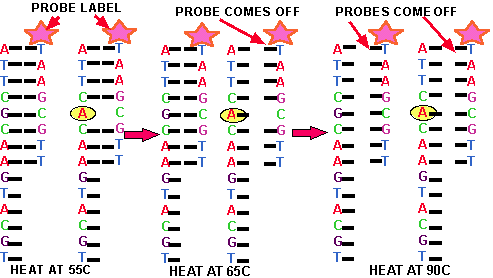
Figure 9. Effect of Temperature on Hybridization. In this figure a short piece of DNA, called the PROBE (Pink Star) is mixed with two different long DNA strands. One of these long strands contains a sequence of base pair that exactly matches that of the probe, whereas the other differs with respect to a single base pair (yellow oval). At 55oC the probe binds to BOTH long DNA molecules, however at 65oC the probe does not bind to the long DNA molecule with a single base pair mismatch. The probe is unable to bind to either long DNA molecule at 90oC. What do you think might be the results at a temperature of 60oC?
![]()
Hybridization is carried out as illustrated in Fig. 10:
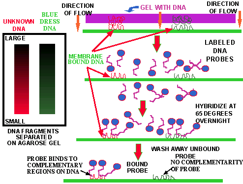
Figure 10. The hybridization procedure. The DNA samples are cut with R.E. and the fragments separated on a gel (left). The gel is placed on top of a DNA-binding protein and the DNA-fragments are washed directly onto the membrane where they stick. A short piece of DNA (the PROBE), to which a reporter molecule is attached, is heated with the bound DNA overnight. During this time the probe will bind to any complementary DNA attached to the membrane. All the unbound probe is subsequently washed away. The reporter is visualized so the location of the sample DNA fragments complementary to the probe DNA can be seen.

Summary animation of the first step in hybridization; transfer of DNA from agarose gel to membrane. For a professional animation of Southern Blotting that requires Shockwave/32MB memory/Pentium processor click hear and load the appropriate file.

Summary animation of hybridization procedure (Southern Blot); probing of DNA bound to membrane.
The entire procedure is called HYBRIDIZATION. Hybridization works because of the SEQUENCE OF THE PROBE. A given probe will only bind with DNA that is attached to the membrane IF IT FINDS a MATCHING or COMPLEMENTARY DNA base pair sequence that forms enough bonds to remain together under the heating conditions used. Thus if no matching complementary sequences are found, NO PROBE will bind (Fig. 10, green DNA). If one or two of the 1,000s of the bound DNA fragments contain a sequence complementary to the sequence in the probe, the probe will BIND only to these fragments and thus become ATTACHED TO THE MEMBRANE through this association with the complementary membrane-bound-DNA (Fig. 10, red DNA). It is possible to chose probes that are known to bind to specific sequences or artificial probes of KNOWN SEQUENCES can be made, for about $1.00/base and sent to you in 48 hr.; THEY CAN BE ORDERED OVER THE INTERNET.
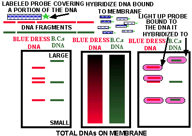
Figure 11. Results of Probing the Separated Fragments of Two DNA Samples. The original gel, shown in the middle, produces a smear of genomic DNA fragments (see #Fig. 13). Within those 1000's of fragments are the fragments shown on the left gel cut from a section of the two original DNA molecules shown in the upper left. When the smear of fragments, fixed to a membrane, are hybridized with the probe (DNA with attached reporter [green star], Fig. 8), the probe will HYBRIDIZE (bind) only to those fragments containing sequences of DNA that complement sequences present in the probe; i.e., those regions DIRECTLY BELOW the probe DNA. The location (pink ovals) of such complementary membrane-bound-fragments are shown on the gel at the right. Thus the detection system only "SEES" the pink ovals. See Fig. 13 for X-ray film detection of DNA fragments from a genomic DNA smear.
In Fig. 12 three different PROBES (A, B, & C) are hybridized against the separated membrane-bound-DNA fragments shown on the right (from 3 duplicate gels). As an exercise determine WHICH BANDS each of the three probes will hybridize (bind) to and light up; that is, draw three duplicate gels, 1, 2, & 3 and circle the band(s) in gel 1 that probe A will bind to; in gels 2 & 3 do the same with probes B & C respectively.
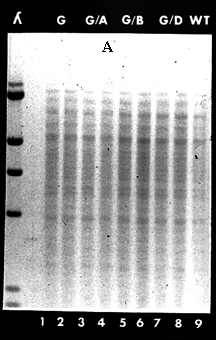
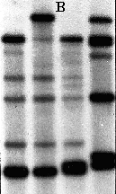
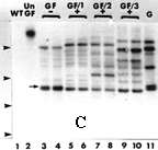
Figure 13. Experimental Gels. These are three experimental gels showing the outcome of a hybridization procedure. Gel A shows the banding pattern seen when genomic DNA, digested with a restriction enzyme, is separated on an agarose gel. The smears in lanes 1 to 9 represent 1,000s of individual DNA fragments produced by the restriction enzyme digestion. The banding seen in these lanes is due to the high concentration of certain genes. Gels B & C show examples of banding patterns obtained when genomic DNA smears, bound to a membrane, are hybridized with specific probes. The probes only bind, or hybridized to, a few of the 1,000s of fragments that contained base sequences COMPLEMENTARY to those on the probe. Attached to the probes is substance which produces LIGHT when treated with certain chemicals and this light is detected with photographic film, in effect taking a picture of each of the locations where the probes bind to a complementary DNA fragment on the membrane. click here and then click on "Hybridization" for some other cartoons of Southern Blotting and illustrations of other molecular biology techniques. Click here for another illustration of hybridization.
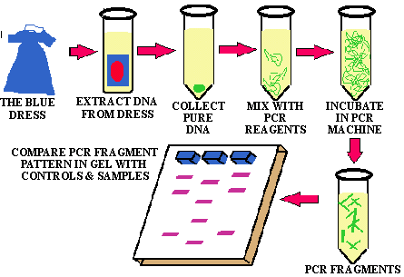
Figure 14 illustrates how the stain from Monica's blue dress was tested to determine if it contained the President's DNA. The DNA from the stain was extracted, digested with one or more restriction enzymes, amplified by the PCR technique and separated on gels. The separated fragments were transferred to DNA-binding membranes and hybridized with various human-DNA probes. The pattern of the DNA fragments (bands) in the various samples that "light up" were then compared for similarities and differences. Recently it was reported that whatever a person handles retains some of that person's DNA. For example, if you pick up a glass or a cup or a shirt or touch a door knob you or lick a stamp you leave some of your DNA behind which can be used to identify you. Even wiping off your "fingerprints" may not remove the DNA totally.
FAQ: About DNA fingerprinting.
1 How good (accurate) is it at identification. For example, is it as good as classical fingerprints?
Answer: In theory, with the exception of identical twins, EVERYONE on this planet has a different DNA fingerprint. That is, DNA fingerprinting IS as good (distinctive/unique/specific) as classical fingerprinting for identification.
2. What are its advantages?
Answer: In theory DNA fingerprinting will work with much smaller amounts of material than a classical fingerprint & DNA lasts much longer than classical fingerprints. DNA-containing samples that are many years old (up to 25 million yr.) are still usable. Only very tiny quantities of DNA are required in order to carry out a highly accurate test. For example, dried blood, semen, spit, skin etc. on samples stored in dusty files for years are still usable. Samples of mixed DNA's can also be used. DNA containing evidence is much harder to clean up at a crime scene than other evidence, like classical fingerprints.
3. What are its limitations?
Answer: There currently are no accepted Federal standards for controlling the quality of DNA testing nationwide. Poor quality & poorly controlled testing leads to QUESTIONABLE and SHODDY RESULTS. Lab A may use one set of procedures and standards, whereas, lab B may use another set of procedures and standards. Making comparisons between results from the two labs difficult. The quality of laboratory personal is not standardized. For example, clinical laboratory technicians, that work in hospitals and clinical labs, must be certified by their states and by a national organization that sets high standards of training and experience. The clinical labs are frequently tested to determine if their procedures are done correctly. They receive & must analyze test samples whose composition is known by certification agencies. The results are returned to the certification agencies for evaluation. If a clinical lab makes too many errors the lab will be DECERTIFIED. This is not yet the case with DNA testing facilities.
Even if there is a perfect match between DNA, you can not say HOW the DNA containing sample got there or WHEN. In the O.J. trial a VALID question was raised about the possibility of evidence being planted. What makes this charge so powerful is the EXTREME SENSITIVITY of the procedure. That is, it would NOT BE DIFFICULT for someone to collect a small quantity of blood (one or two drops from a tube of a blood sample), body fluid or tissue from a victim (e.g. hair in a comb, blood on a Kleenex etc. ) and place this material where it would incriminate an innocent person. There are documented cases where quantities of drugs, marked money etc. have been "planted" and innocent people convicted of crimes because of these actions.
Finally, blood that is mixed with the wrong chemicals or is degraded is difficult to analyze accurately.
4. When faced with DNA evidence what questions should be asked?
Answer: As indicated above (and in the O.J. trial), questions regarding the handling of the evidence, the quality of the testing, including the controls used, the skill of the testing personnel, the accuracy of the data interpretation, possible contamination of the evidence, the possibility of accidental, or intentional misplacement of evidence are all valid questions that should be raised regarding DNA evidence (or any evidence for that matter). As the sensitivity of this powerful technique improves and its use widens throughout our justice system, it is important that we deal with these problems if we expect EQUAL JUSTICE for all. We must not lose sight of the fact that no matter how powerful a new tool in crime fighting is, its ultimate effectiveness is only as good as the persons using that tool.
For a lecture, with images, on DNA fingerprinting visit this site.
![]()
![]()
The polymerase chain reaction (PCR) won its discoverer a Nobel Prize in 1993. Kary Mullis has recorded the thought process he went through as he conceived of the idea and he states that he knew immediately that he was going to become very "FAMOUS" because of this discovery. Everyone else assumed that he would win the Nobel Prize as soon as they heard of the PCR reaction. The ironic thing about this discovery, is that all the scientific information had been around for approximately 20 years and had been overlooked by the greatest scientific minds in the world. This discovery is now seen as such an obvious concept that some scientists question if Mullis deserved the Nobel Prize for such a "simple" and "obvious" discovery. After you hear the PCR story you can make up your own mind?
The PCR is another one of these discoveries, like that of the structure of DNA, the #transforming factor determining the chemical nature of DNA, and restriction enzymes, that generated a quantum leap in science. Almost the instant after PCR is explained to a biological scientist, ideas as to its uses begin to pour from all but the most dull scientific mind. Even today, years after its discovery, we are still developing new uses for PCR.
The irony of the PCR is that living organisms have been doing it since they #evolved in the primeval organic soup of this planet 3.5 billion years ago and scientists have been aware of the general DETAILS of this process for ~50 years. To put it succinctly, the PCR does in the test tube what every bacterium does in its tube of media or on an agar-plate and each of us do every day; we all produce billions of exact copies of our own DNA; AMPLIFYING our DNA millions of time. The enzyme DNA polymerase was discovered in the 1950s and our knowledge of the process has been increasing ever since. This means that thousands of scientists have studied DNA replication for 40 years without tumbling to PCR.
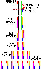
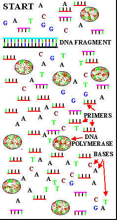
The basic principle of PCR is shown in Fig. 15. It has been know for a long time that DNA polymerase requires a short strand of DNA or RNA called a PRIMER to "prime" the START OF DNA REPLICATION. Mullis's genius was that he reasoned "That if you added the following components to a test tube containing a single DNA molecule, you could replicate & amplify that DNA molecule many million fold in a short time.
The components are:
![]() A sample of the TARGET DNA
to be copied. In theory only a single molecule is needed.
A sample of the TARGET DNA
to be copied. In theory only a single molecule is needed.
![]() A set of short (15 to 40 bases) single stranded PRIMERS
of DNA, in EXCESS,
that will bind to complementary regions of the opposing stands of the TARGET
DNA molecule . These primers
BRACKET the region of DNA to be amplified.
A set of short (15 to 40 bases) single stranded PRIMERS
of DNA, in EXCESS,
that will bind to complementary regions of the opposing stands of the TARGET
DNA molecule . These primers
BRACKET the region of DNA to be amplified.
![]() An EXCESS of the 4 nucleotide triphosphates, ATP, GTP, CTP, TTP.
An EXCESS of the 4 nucleotide triphosphates, ATP, GTP, CTP, TTP.
![]() The enzyme, DNA polymerase.
The enzyme, DNA polymerase.
![]() Various buffers and cofactors like magnesium ions required by DNA polymerase.
Various buffers and cofactors like magnesium ions required by DNA polymerase.
The final trick was to get the two target DNA strands APART (separated) so the primers could bind and the DNA polymerase could do its thing. It had been known for ~50 years that heat separates DNA strands and that complementary strands then rejoin through base pairing when the temperature is subsequently lowered. So Mullis heated his mixture of target DNA, primers and triphosphate nucleotides to about 90oC for a few minutes to separate the target DNA. He then lowered the temperature enough to allow the primers, which were small and in VAST EXCESS, to bind (ANNEAL) to their respective complementary target DNA base pair-sequences. At this point he added DNA polymerase and allowed the polymerization reaction with the triphosphate nucleotides to occur. That is, the DNA polymerase FILLED IN the missing portion of each strand making TWO NEW DOUBLE STRANDED regions of DNA.
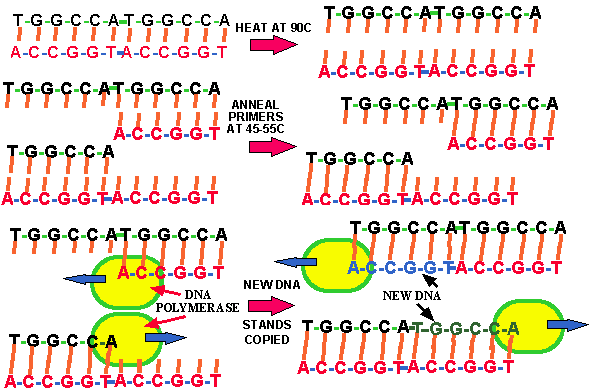
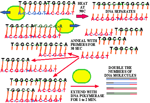
Figure 15. The polymerase chain reaction. For another figure illustrating the PCR click here and view the "PCR" link. Click here and here for brief discussions of PCR.
Summary animation of PCR. For a professional animation that requires Shockwave/32MB memory/Pentium processor click here and load the appropriate file.
Each entire PCR cycle takes only 2 to 10 minutes. However, there was one problem with the system devised by K. Mullis, which was that the DNA polymerase he used was DESTROYED BY THE HEATING process. Mullis had to add new DNA polymerase for EACH ROUND, which was time-consuming and expensive. This problem was solved by using HEAT-RESISTANT DNA polymerase that only needed to be added once. #THERMOPHILIC bacteria that live in boiling hot springs have been described previously. This is another case of SERENDIPITOUS BASIC SCIENCE turning out to be important. The study of thermophiles might seem to be a waste of money and time to many people as these bacteria, while certainly interesting, don't cause disease in man or any other life form. The argument could be made that a scientist could better spend his/her time working on cancer or some other terrible disease that afflicts humankind. However, it turns out that since thermophilic DNA polymerases tolerate high temperatures (e.g. 90oC) for long periods without being destroyed, they are the perfect solution to the PCR DNA polymerase problem. Thus today PCR has become a revolutionary tool because of scientists who studied these odd thermophiles.
|
|
The standard PCR reaction is run through about 30 cycles in a couple of hours which results in the amplification of the original DNA by over a 109 fold. Thus a single specific DNA region can be amplified to yield sufficient quantities to do anything that can be done with bulk-isolated DNA. In the case of crime evidence this means that the DNA in a single hair follicle, a single drop of semen or blood is sufficient to prove that an individual was present at the scene of a crime. Indeed any piece of evidence that contains one or more moderately long DNA fragments (e.g. >200 base pairs) can be amplified with PCR and its RFLP determined for identification purposes---or maybe to build a dinosaur eventually. However, at present the maximum limit for amplification is approximately 42 kilobases (42,000).
![]()
![]() DNA fingerprinting is a powerful technique that is capable of
distinguishing every individual organism from every other individual organism on
this planet with the EXCEPTION of identical twins or clones.
DNA fingerprinting is a powerful technique that is capable of
distinguishing every individual organism from every other individual organism on
this planet with the EXCEPTION of identical twins or clones.
![]() The major limitations of DNA fingerprinting are human error and the quality of
the technology used.
The major limitations of DNA fingerprinting are human error and the quality of
the technology used.
![]() The discovery of PCR increased the sensitivity and versatility of DNA
fingerprinting, and changed the face of molecular biology.
The discovery of PCR increased the sensitivity and versatility of DNA
fingerprinting, and changed the face of molecular biology.
![]() These techniques will allow infectious diseases to be identified within hours or
even minutes. They are accelerating the rate of sequencing the human genome,
they allow the investigation of complex ecological interactions and species
relationships that here-to-fore have been too complex to study.
These techniques will allow infectious diseases to be identified within hours or
even minutes. They are accelerating the rate of sequencing the human genome,
they allow the investigation of complex ecological interactions and species
relationships that here-to-fore have been too complex to study.
WELCOME TO THE NEW WORLD OF GENETIC ENGINEERING; JUST KEEP YOUR SEAT BELTS FASTENED BECAUSE IT IS GOING TO BE A WILD RIDE.
![]()
Signal transduction refers to the most common method of regulation of complex cell activity. It involves an external signal molecule (ligand) being bound by a specific cell's membrane receptor. The receptor, in turn triggers changes in the internal command system of the cell causing it to undertake a new activity so long as the ligand is present. A powerful new therapy using STGT is being developed whereby part of the genome of a common cold virus is replaced with:
In this procedure the patient is infected by the modified virus. The modified viral DNA then remains in the patient's cells. The therapy gene remain inactive until the "triggering" substance was given externally. The amount of triggering substance would determine the degree to which the therapy gene was turned on (like a dimmer switch).
![]()
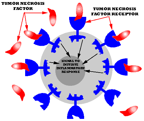
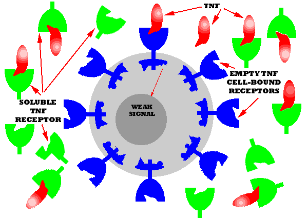
A cytokine known as the TUMOR NECROSIS FACTOR (TNF) is known to be responsible for much of the effects of rheumatoid arthritis. It induces immune cells to produce substances that attack the host's own body cells causing the painful inflammation that accounts for most of the problems of rheumatoid arthritis. The TNF acts by binding to a specific membrane-bound protein receptor on the immune cells. The receptor protein, which extends into the cell cytoplasm undergoes a change in structure which causes it to react with other proteins within the cytoplasm which sets up a signal cascade that ends in the translation and transcription of the genes producing and releasing the damaging material. Using GE technology a truncated (shortened) form of the receptor gene has been cloned. This gene makes a soluble form of the receptor that contains the TNF BINDING SITE. When large quantities of the material is injected into an arthritis patient it binds the TNF thus preventing it from binding to the cellular-associated receptor thus preventing (or minimizing) the signal from being sent.
|
|
|
|
![]()
![]()
Copyright © Dr. R. E. Hurlbert, 1999.
This material may be used for educational purposes only and may not be
duplicated for commercial purposes.