![]()
Pharynx
Author: Dr.R.Menaka
![]()
...........................................................................................................................................................................................
The pharynx is a musculo-membraneous
sac which communicates both
the digestive and respiratory tract.
It is somewhat funnel or conical shaped and situated behind the soft palate,
posterior nares and isthmus faucium.
The cavity of pharynx communictaes by seven openings.
1. Two opening of eustachian tube
2. Two opening of posterior nares
3. Aditus laryngis- one
4. Aditus esophagi -one
5. Isthmus faucium - one
The wall of the pharynx consists of pharyngeal muscles, aponeurosis and
mucous membrane.
The mucous membrane lines the cavity of pharynx and continues with mucous
membrane
of the mouth, nasal chambers, larynx, eustachian tubes and esophagus.
Blood supply- receives from common carotid artery, external maxillary artery and
external
carotid arteries.
Nerve supply-From the pharyngeal plexus, which is formed by the pharyngeal
branches from the
glosso-pharyngeal nerve and the superior cervical ganglion.
Note: Closure of isthmus faucium is brought by the two levatores palati,
which pull the soft palate simeltaneously with the palato-pharyngeus. Thus, the
pharynx is converted into
respiratory type from the digestive type.
Hore: Pharynx is longer and narrower.
The superior wall of the pharynx is related with the guttural pouch in addition.
Esophagus
The esophagus is musculo-membraneous tube which extends from the pharynx
to the stomach.
Its length is about 0.75 to 1 meter.
It divided into cervical and thoracic course.
The cervical part begins from the pharynx to the thoracic inlet.
The thoracic part starts from the thoracic inlet to the hiatus esophaeus.
Passing through this opening, it gains the abdominal cavity and immediately
terminates on the dome
like, rumino-reticular wall.
Stomach
Ox:
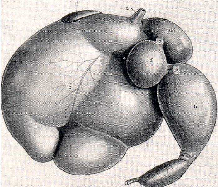
The stomach of the ox is very large and occupies nearly three fourth of the
abdominal cavity.
It completely fills up the left of the abdomen except for a space intended for
the spleen and
extends considerably into the right half. It has four compartments:
Rumen (Paunch); Reticulum (honey comb); omasum (omasal laminae) and abomasum
(true stomach).
Capacity: Varies according to the age, size, breed etc. Medium size 120 litres
to 160 litres.
In the new born the first and second are about half of the fourth.
At 10 to 12 weeks the ratio is reversed.
At four months first and second are 4 times as large as 3rd and 4th put
together.
At 11/2 years omasum equals abomasum.
In the adult, capacity of rumen 80 % , reticulum 5 %, omasum 7-8 % and abomasum
7-8 %.
The Exterior of Stomach
Rumen:
The rumen occupies most of the left half of the abdomen and extends considerably
over the median
plane to the right. It extends from the lower part of the 7th and 8th
intercostal space to the
pelvic inlet. It is slightly compressed laterally and presents, 2 surfaces, 2
borders and
2 extremities. The parietal surface is convex and is related to the diaphragm,
left wall to
abdomen and spleen. The visceral surface is irregular and is related to the
omasum, abomasum,
intestine, liver and pancreas, left kidney, adrenal, aorta and posterior
venacava.
The dorsal curvature is convex an dis in contact with the dorsal wall and
attached to it
by peritoneum and connective tissue as far back as fourth lumbar. The ventral
curvature is also
convex and is related to the floor.
The two surfaces are marked by right and left longitudinal grooves and dividing
it into dorsal and
ventral sacs. The anterior extremity is divided ventrally by a transverse groove
into two sacs- dorsal
and ventral, of which the dorsal one becomes continuous with the reticulum,
curves over the ventral sac,
which is rounded and blind. The junction of the dorsal sac and reticulum is
marked by a groove, the
rumino-reticular groove which is distinct ventrally the two compartments form a
sort of dome-
the atrium ventriculi on which the esophagus terminates. The posterior extremity
extends to the pubis
and is related to the intestine and bladder. It is divided by the deep posterior
transverse groove
into dorsal and ventral sacs both of which are blind.
The grooves lodge the vessels and nerves of rumen.
Reticulum:
The reticulum is the most anterior and smallest of the four compartments. It
extends from the 6th
to the 8th ribs. The greater part of it is to the left of the median line.It is
compressed from
before backwards. The parietal surfaces forwards, convex and lies against the
diaphragm and liver.
The visceral surface faces backwards, is flattened and ends dorsally by joining
the rumen, the concave
line of junction corresponding to ridge in the interior which forms the lower
margin o fthe rumino
-reticular orifice. The lesser curvature faces to the right and dorsally and is
connected with the omasum.
The greater curvature faces to the left and in ventral. The right extremity
forms a rounded blind sac
which is in contact wiht the liver omasum and abomasum and lies opposite to the
sixth intercostal space.
Omasum:
The omasum is ellipsoidal in form and is somewhat compressed between the two
surfaces. It is very clearly marked
off from the other compartments and lies to the right of median line from the
7th to the 11th rib.
The parietal surface to the right and forwards and is related to the diaphragm
and liver. The visceral
fcaes to the left end backwards and is in contact with right face of rumen,
reticulum and abomasum.
The greater curvature faces backwards and to the right. The lesser curvature
faces backwards and to
the right. The lesser curvature is very short and faces forward and to the left.
It is connected at its upper part
with the reticulum. Below it joins the abomasum.
Abomasum:
The abomasum is an elongated sac which lies on the abdominal floor from the
xiphoid cartilage backwards.
The anterior blinded is at the xiphoid region in relationb wiht the reticulum.
The boby extends
back between the ventral sac of rumen and the omasum and turns to the right
behind the omasum.
It is constricted about the middle forming an anterior larger part and a
posterior pear shaped smaller part.
The pyloric part inclines dorsally and joins the duodenumat at the ventral part
of the 10th rib.
The parietal surfcace is in contact with the abominal floor. The visceral face
is related to the rumen
and omasum. The greater curvature gives attachment to the superifical part of
the greater omentum. The
lesser curvature is related to the greater curvature of the omasum.
The Interior of Stomach
Rumen:
The cavity of the rumen is divided into two sacs by the pillars of the rumen
which are muscular folds
and corresponds to the grooves on the exterior. They project like shelves into
the cavity of the organ.
The anterior pillar has a thick concave free edge. Between these free edges, the
two sacs of rumen communicate with each
other. The right and left pillars connect the anterior and posterior pillars and
are less prominent.
The rumino- reticular fold corresponds to the rumino-reticular groove. Its free
edge is concave and
forms the ventral and lateral margins of the large oval rumino reticular
aperture. The cardia is about 10 to 12 cm. ventral
to the vertebral end of the 8th or 9th rib. The opening is slit like. The mucous
membrane is brown
in color except on the pillars where it is pale. It is thickly studded with
papillae which are however
not present on the pillars.
The reticular or esophageal groove begins at the cardia and passes ventrally on
the internal face of the
right wall of the atrium and reticulum to end at the reticulo-omasal orifice. It
is about 18 to 20 cm length.
Its direction is chiefly dorso-ventral but usually it inclines somewhat forward
and medially in its
ventral part. The groove is twisted in spiral fashion so that its thickened
edges project at first backward
then to the left and finally forwards. The twist mainly concerns the left lip.
Reticulum:
The interior of the reticulum is raised into folds about 1/2 inch high enclosing
4 to 6 sided spaces or cells (honey comb).
These cells are subdivided by smaller folds and bottoms are studded with pointed
horny papillae. The reticulo-omasal orifice is
situated at bthe lesser curvature of the reticulum five or six inches above the
bottom of the latter and is rounded.
Omasum:
The cavity of the omasum is occupied by about hundred longitudinal muscular
folds-the laminae omasi which spring from the greater curvature.
The largest of these about half a dozen in number have a superior convex
attached edge and a thick concave free egde. A groove sulcus
omasi extends from the reticulo-omasal opening to the omaso-abomasal opeining
and is about 4 inches long. It is free from
lamina. The omaso-abomasal orifice is oval and is about 4 inches long.
Abomasum:
The cavity of the abomasum is divided into two parts by a constriction. The
first part is lined with
soft glandular epithelium forming about a dozen or more spiral folds. The second
part is narrower,
and pear shaped and presents a brownish mucous membrane. the pylorus is small
and round.
Blood supply:
Right and left ruminal, reticular and omaso-abomasal arteries.
Nerve supply:
Vagus and symnpathetic (coeliac plexus).
Horse:
It is simple sharply curved U-shaped sac, the right part being shorter than the
left. Capacity is 2 to 4 gallons(8-16 litres). It is situated in the left
dorsal part of the abdominal cavity behind the diaphragm and liver mainly to the
left of the median line. The parietal surface faces forwards
upwards and to the left and is related to the diaphragm and liver. The visceral
surface faces backwards, downwards
and to the right and is related to the colon and small intestine. The lesser
curvature is short. The greater curvature
is extensive and is related on the left to the spleen and the rest to the great
colon. The left dorsal extremity
or saccus caecus lies ventral to the dorsal end of 16th or 17th rib. It is
related to the pancreas and terminal part of the
greater colon behind and the base of the spleen laterally. The right extremity is smaller and is continued by the
duodenum. It is attached by
1. Gastrophrenic ligament
2. Greater omentum
3. Gastrophrenic omentum
4. Lesser omentum
5. Gastrophrenic fold.
Interior:
The mucous membrane is divided into two parts by a sinous ridge-margo plicatus.
That part on the left
extremity is non-glandular being the extension of the esophageal mucous
membrane-esophageal region and
the remaining is glandular. The glandular part is subdivided into three zones,
according to the type of
glands present, but no distinct line of demarcation exists. A narrow zone along
the margo-plicatus is the
cardiac gland region. Adjacent to it is the large fundic gland region. Remainder
of the mucous membrane
is the pyloric gland region.
Dog:
Capacity is about 3 litres.
When full it is pyriform.
The left part is large and rounded while the right part is small and forwards,
downwards and to the left. It is
related to the liver; diaphragm and left ventral and lateral abdominal wall as
far as the level of the
level of the 2nd or 3rd lumbar.
The visceral surface is less extensive and is related to the intestine, pancreas
and left kidney.
The lesser curvature is nearly straight above but below it makes a sharp bend
forming an angle.
The greater curvature is extensive and it extends, when the stomach is full
behind the costal arch.
Ventrally it lies on the abdominal floor about midway- between xiphoid cartilage
and pubis.
Fowl:
It is made up of two parts. Proventriculus (glandular) and gizzard (muscular).
The proventriculus is an elongated fusiform thin walled tubular organ, related
laterally and ventrally to the liver and the spleen
at its superioposterior aspect. It is connected in front with the esophagus and
behind with the gizzard. Isthumus
is lined by glandular columnar epithelium.
The glandular or muscular stomach is a thick walled muscular disc with two
orifices placed close together on the antero-dorsal aspect of its circumference.
It is situated behind and party between the two lobes of the liver. The mucous
membrane, lining the gizzard
is thrown into ridges and is covered by dense horny subsatnce, secreted by the
glands lying
beneath the columnar epithelium.