 The
nucleus contains most of the genes that control the eukaryotic cell
(some are located in the mitochondria). When viewing a cell through a
microscope, the nucleus is the most obvious and visible organelle,
averaging 5 micrometers in diameter.
The
nucleus contains most of the genes that control the eukaryotic cell
(some are located in the mitochondria). When viewing a cell through a
microscope, the nucleus is the most obvious and visible organelle,
averaging 5 micrometers in diameter.The
nucleus contains most of the genes that control the eukaryotic cell
(some are located in the mitochondria). When viewing a cell through a
microscope, the nucleus is the most obvious and visible organelle,
averaging 5 micrometers in diameter.
The nuclear envelope separates the nuclear contents from the cytoplasm. It is a double membrane, composed of two lipid bilayers that are separated by about 20-40 nm of space. The envelope is perforated by pores about 100 nm in diameter which allow material to move in and out of the nucleus. The membranes of the nucleus fuse at the pore lips. Protein structures called pore complexes line each pore, regulating the flow of macromolecules and particles. The internal side of the nuclear membrane is lined with a web-like formation of protein filaments, called the nuclear lamina, that holds the shape of the nucleus. There is evidence of a nuclear matrix--a framework of fibers extending throughout the inside of the nucleus.
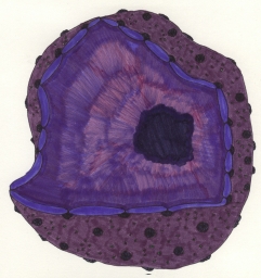
Inside the nucleus is the
nucleolus, roughly spherical-shaped, which appears under a microscope
to be a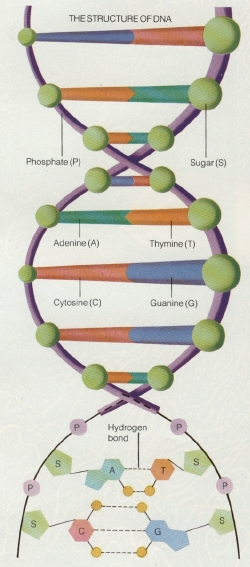 mass of dense granules and fibers. In the nucleolus components of
ribosomes are synthesized and assembled, and then pass through the
nuclear pores. Once in the cytoplasm, they combine and form
ribosomes.
mass of dense granules and fibers. In the nucleolus components of
ribosomes are synthesized and assembled, and then pass through the
nuclear pores. Once in the cytoplasm, they combine and form
ribosomes.
The nucleus controls protein synthesis in the cytoplasm by sending out mRNA (messenger ribonucleic acid). The mRNA is synthesized in the nucleus as instructed by the DNA and sends genetic messages through the pores and attaches to ribosomes. The genetic message that the mRNA carries is translated into the basic structure of a specific protein.
DNA stays inside the nucleus, and RNA can leave. RNA transcribes from the DNA, beginning at a promoter (a specific sequence that binds to RNA polymerase and tells the RNA where to begin transcribing). A terminator tells the RNA where to stop. As the RNA leaves the nucleus, the promoter and terminator come off.
There are two "sections" of DNA: exons and introns. Exons are sequences that actually code for proteins, and introns are "junk DNA" that do nothing except hold exons together.
In the diagram at right, the structure of DNA is shown in three different ways. The top section separates DNA into phosphate (the purple segments), sugar (the green balls), and nitrogenous bases (the green, orange, blue, and red 'rungs'). The middle section identifies the the four nitrogenous bases present in DNA: adenine, guanine, cytosine, and thymine (uracil takes its place in RNA). The bottom section is more complex, showing the structure of the DNA components and the bonds between the nitrogenous bases.