The
Extracellular Matrix (ECM)
 The
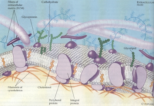
extracellular matrix is composed primarily of glycoproteins (proteins
covalently bonded with carbohydrate, usually short chains of sugars)
secreted by cells. The most abundant glycoprotein in the ECM is
usually collagen which forms strong fibers outside the cells (the
striped purple structures at left). These collagen fibers are
embedded in a web of proteoglycans--molecules especially rich in
carbohydrates (up to 95%) and can form large complexes (the blue,
willowy structures). Another glycoprotein, fibronectin, binds to
receptor proteins called integrins that built into the plasma
membrane (which is about 0.00001 millimeters thick). Integrins span
the membrane and bind to microfilaments on their cytoplasmic side,
and are able to transmit between the ECM and the
cytoskeleton.
The
extracellular matrix is composed primarily of glycoproteins (proteins
covalently bonded with carbohydrate, usually short chains of sugars)
secreted by cells. The most abundant glycoprotein in the ECM is
usually collagen which forms strong fibers outside the cells (the
striped purple structures at left). These collagen fibers are
embedded in a web of proteoglycans--molecules especially rich in
carbohydrates (up to 95%) and can form large complexes (the blue,
willowy structures). Another glycoprotein, fibronectin, binds to
receptor proteins called integrins that built into the plasma
membrane (which is about 0.00001 millimeters thick). Integrins span
the membrane and bind to microfilaments on their cytoplasmic side,
and are able to transmit between the ECM and the
cytoskeleton.
Membrane proteins serve many
functions, including transport, enzymatic activity, signal
transduction, intercellular joining, cell-cell recognition, and
attachment. A protein that spans the membrane may provide a
hydrophilic (water-loving) channel across the solute-selective
membrane. Some transport proteins hydrolyze ATP to actively pump
substances across the membrane. A protein built into the membrane may
be an enzyme, active site exposed to substances outside of the cell.
Sometimes, enzymes are present in a series, carrying out sequential
steps in a metabolic pathway. Membrane proteins may have a binding
site with a specific shape that fits a chemical messenger, like a
hormone. The signal may change the conformation of the protein that
relays the message to the inside of the cell. Membrane proteins of
adjacent cells may be hooked together in various ways. Some
glycoproteins work as identification, recognized by other cells.
Also, fibers may be bonded to membrane proteins, which helps the cell
maintain its shape and fixes the location of certain membrane
proteins. Proteins adhered to ECM can coordinate extracellular and
intracellular changes.
 The
animation at right illustrates the fluid mosaic model. Originally, it
was theorized that the phospholipid bilayer was sandwiched by sheets
of proteins, but now the working model is one where proteins move in
and around the bilayer. The phospholipids themselves have hydrophilic
heads and hydrophobic tails. The proteins also have hydrophilic and
hydrophobic regions which correspond with the
phospholipids.
The
animation at right illustrates the fluid mosaic model. Originally, it
was theorized that the phospholipid bilayer was sandwiched by sheets
of proteins, but now the working model is one where proteins move in
and around the bilayer. The phospholipids themselves have hydrophilic
heads and hydrophobic tails. The proteins also have hydrophilic and
hydrophobic regions which correspond with the
phospholipids.