The
Cytoskeleton
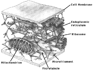 A
network of fibers exists throughout the cytoplasm, supporting the
cell and maintaining its shape--a function that is particularly
important in animal cells, which have no cell wall. In addition to
stabilizing the cell, the cytoskeleton provides anchorage for
organelles even cytosolic enzyme molecules.
A
network of fibers exists throughout the cytoplasm, supporting the
cell and maintaining its shape--a function that is particularly
important in animal cells, which have no cell wall. In addition to
stabilizing the cell, the cytoskeleton provides anchorage for
organelles even cytosolic enzyme molecules.
The cytoskeleton is also
involved several types of cell motility, ranging from changes in cell
location to movements of certain cell parts. This generally requires
an interaction between the cytoskeleton and proteins called motor
molecules, which power cilia and flagella, and also cause muscle
cells to contract. Vesicles may travel on cytoskeletal "tracks". The
cytoskeleton also manipulates the plasma membrane to form food
vacuoles during phagocytosis. There is evidence that cytoskeleton may
be able to transmit mechanical forces from the cell's surface to its
interior, perhaps even into the nucleus.
There are three main types of
fibers: microtubules (the thickest of the three), microfilaments (the
thinnest), and intermediate filaments (middle range, as the name
suggests).
MICROTUBULES
These fibers are found in the
cytoplasm of all eukaryotic cells. They are straight, hollow rods
measuring about 25 nm in diameter, and from 200 nm to 25 micrometers
in length. The wall of the tube is made from a globular protein
called tubulin. Microtubules elongate by adding tubulin molecules to
the ends, and can be disassembled and their tubulin used to build
other microtubules elsewhere.
 They
shape and support the cell, serving as tracks which organelles with
motor molecules can move along.
They
shape and support the cell, serving as tracks which organelles with
motor molecules can move along.
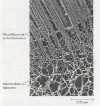
MICROFILAMENTS
Unlike microtubules,
microfilaments are solid robs, approximately 7 nm in diameter. They
are a twisted double chain of subunits of the globular protein actin.
They seem to be present in all eukaryotic cells, bearing tension. In
combination with other proteins, they often form a three-dimensional
network just inside the membrane, supporting the cell's shaping and
giving the outer cytoplasmic layer a slightly more solid consistency
than the inner cytoplasm.
INTERMEDIATE FILAMENTS
The diameter of these
tension-bearing filaments ranges from 8-12 nm. This is a very diverse
class of cytoskeletal elements, each type constructed from different
families of proteins. Intermediate filaments are more permanent than
the other fibers. Chemical treatments that would disassemble the
others do not remove these filaments, which retain the original shape
of the cell. A cage of intermediate filaments usually surrounds the
nucleus.