


Patient Selection
An FHO is most successful in small dogs (those less than 40-50 lbs.) and cats. Larger dogs may have success with the procedure, but it is less predictable because the resulting "false joint" is not as stable as a true bony joint. This instability makes the new "joint" less able to handle the stresses of the heavier animal. For larger animals, a total hip replacement may be a better choice, depending on the animal's pathology and the owner's financial means.
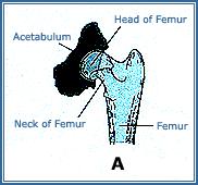

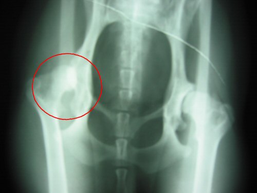
In simple terms, an FHO involves cutting off the head ("ball" portion) of the femur at a steep angle to remove the bony connection between the femur and the pelvis. This eliminates the hip joint as well as the pain associated with bone-on-bone movement.
In a normal hip joint, both the femoral head and the acetabulum of the pelvis (see diagram above) are covered in smooth cartilage which cushions and protects the two bones during joint motion. In animals with pathology of the hip joint, this cartilage is either eroded or otherwise no longer protecting the area where the two bones come into contact. When the pelvis and femur contact each other directly and rub against one another, pain results. By removing bone, surgeons can eliminate this painful bony contact resulting in more comfortable movement for the patient.
Intuitively, one wonders how the hip continues to function after a surgery which basically eliminates the joint. After the procedure, fibrous scar tissue forms between the new end of the femur and the pelvis, effectively creating a new soft tissue barrier with which to cushion both bones during movement. The thigh muscles are strong enough to continue to move the leg in a normal fashion while the fibrous tissue acts as a new "false joint".

Recovery following an FHO is different from other orthopedic procedures. Instead of having the animal strictly confined to cage-rest for a prolonged period, the goal is to get up and get moving! The first 7-10 days following the surgery, the animal is restricted in his movement, protected from slippery floor surfaces, and not allowed to over-do exercise. The owner is responsible for passive range-of-motion physical therapy where the leg is pushed and pulled through normal movements. The pet ofted begins toe-touching to bear small amounts of weight within days after the surgery.
After the first 1-2 weeks, the sutures are removed and the animal is encouraged to walk, run, swim and play. This activity is important because the animal naturally places weight on the leg (which helps fibrous tissue form) and puts the leg through range-of-motion exercises which helps prevent too much scar tissue from forming in inappropriate places that could limit use and motion of the leg long term.
Long Term Prognosis
Lighter animals who quickly go back to moderate to full activity usually have a good long-term prognosis for comfortable use of the leg. Because a bony joint no longer exists, arthritis of the area is not a usual sequella of this surgery as it can be in other orthopedic procedures. Heavier animals may have a less stable joint because of the excess joint strain and discomfort may be associated with this instability.

(2)
(3)
(4)
