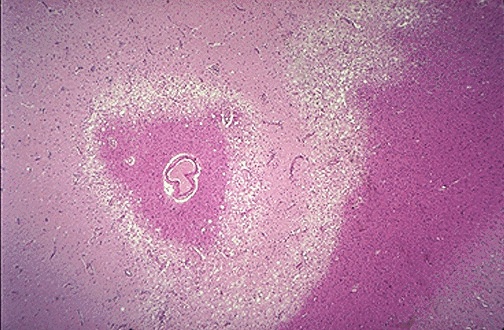
Brain Infarct
Click to see other slides: [1]
1.
What type of tissue necrosis is shown?
Liquefactive necrosis.
2.
What are the characteristic histopathologic features?
No residual tissue architecture is preserved, pink staining cellular debris,
large number of macrophages.
3.
What is the underlying cause of this pattern of necrosis?
Cerebral hypoxia within the CNS.
May also be due to focal bacterial or occasionally fungal infections.
4.
Why does it result in this particular appearance in brain tissue?
White matter contains myelin and myelin contains a high lipid content which
is rapidly dissolved.
It is also due to the relative lack of extracellular structural
protein (reticulin and collagen), which leads to a rapid loss of tissue
architecture.
5.
Give some possible clinical manifestations of this condition if the
affected area was located in the region of the left internal capsule?
There is motor paralysis of the limbs because internal capsule contains the
corticospinal tract.
There is also paralysis of some axial muscles, but this may be transient
because there are other tracts carrying out the same function which is
unaffected.
6.
Is this reversible and why?
No, this is not a reversible process.
Neurons are permanent cells and cannot be replaced.
There is astrocytosis because of astrocyte proliferation.
7.
What is the long-term naked eye appearance of this area?
An empty cavity will be formed in place of the lesion.
<< PREVIOUS INDEX NEXT SLIDE >>
Copyright � Joseph Ong 2003