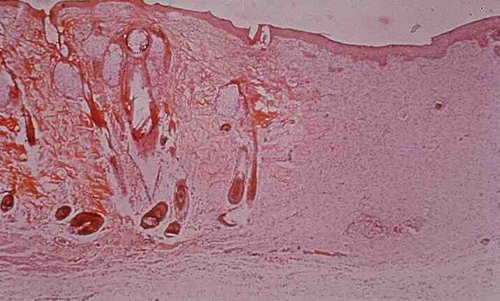
Skin: Granulation tissue
1.
What pathological process is illustrated now?
Granulation
formation, healing
2.
What
type of tissue is present in the a) initial stages and b) later stages?
a)
Endothelium of capillaries, inflammatory cells (macrophages,
lymphocytes), fibroblasts (vascular granulation tissue)
b)
Collagen, fibroblasts (fibrous granulation tissue)
3.
Describe
the characteristic histopathological features in this slide.
Many
new capillaries perpendicular to the surface in the granulation tissue.
Some inflammatory cells present, but it is normal, may not be signs of
infection. Some
neutrophils, dead cells where the epidermis should have seen.
Area filled with fibroblasts.
4.
Number
the following pathophysiological processes involved in would healing according
to sequence:
1.
Angiogenesis
2.
Migration and proliferation of fibroblasts
3.
Deposition of extracellular matrix
4.
Tissue remodelling
5.
What�s
the difference btw healing by 1st and 2nd intentions?
What does this case show?
1st
intention: wound
edges in apposition, wound completely healed by 2nd month
2nd
intention: arge
tissue defect with blood cot and tissue debris.
Abundant granulation tissue grows to fill the wound.
Wound contraction caused by myofibroblasts, so defect markedly reduced
from original size. Organisation
and filling by granulation tissue take considerably longer.
6.
What will be the outcome in this case wrt the states of the a) epidermis, b)
skin appendages, c) dermis, d) deeper soft tissue
a)
reepithelisation � proliferation of epithelium at edges of the defect
b)
Skin appendages (sweat glands, sebaceous gland) lost permanently
c)
dermis interrupted by scar tissue
d) infiltration with scar tissue
7.
What
factors can delay the healing process in this case?
Systemic
factors:
- nutritional
status (protein and vit. C)
-
metabolic status (diabetes delay
healing)
-
adequacy of blood supply
-
hormones (glucocorticoid therapy
hinders inflammatory-reparative process)
Local
factors:
-
infections: presence of
inflammatory cells delay healing.
-
mechanical factors: increased
abdominal pressure may rupture abdominal wounds.
-
foreign bodies: presence of
foreign bodies induce a giant cell reaction - continued presence of inflammatory
cells leads to delayed healing.
-
type of wound: wounds with large
defects take longer to heal.
8.
When
fully healed, skin wounds usually have tensile strength 70-80 % that of
unwounded skin.
<< PREVIOUS INDEX NEXT SLIDE >>
Copyright � Joseph Ong 2003