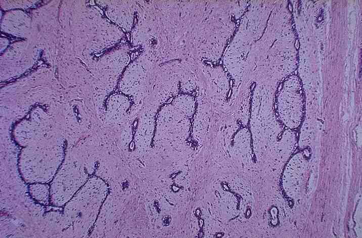
Breast: Fibroadenoma
A
30 yr old woman felt a 1.5cm diameter painless, firm, mobile nodule in her
breast.
1.
What are differential diagnoses in order of likelihood in this young woman?
Fibrocystic
changes
Fibroadenoma
Ductal
carcinoma
2.
What are the pathological features (macroscopic, microscopic) that make this a
benign tumour?
Macroscopic:
well circumscribed, mobile, not attached to underlying structures, surrounding
skin is not ulcerated or edematous.
Microscopic:
Intact fibrous capsule and lobular structure. Glandular cystic spaces
lined by epithelium, surrounded by loose cellular, myxomatous stroma
No
pleomorphic nuclei, mitoses
Compressed
ducts regularly spaced
3.
Name the histological features of this lesion.
They consist of stroma and epithelium lining cystic spaces compressed by the stroma.
4.
What are the methods available for obtaining samples for tissue confirmation
(i.e. histological diagnosis)
Fine
needle aspiration
5.
What is the corresponding imaging appearance (mammogram, ultrasound)?
Will
circumscribed density, more dense than surrounding tissue
No
calcification
No
/ minimal distortion
(compare
with �stellate� mammogram)
Note:
for cancerous breast lesions, pleomorphic calcification �
bad prognosis
6.
What is the treatment of choice?
Surgical
excision
7.
What is the risk of
Recurrence?
Malignancy? Associated with a small increase in risk of breast cancer
<< PREVIOUS INDEX NEXT SLIDE >>
Copyright � Joseph Ong 2003