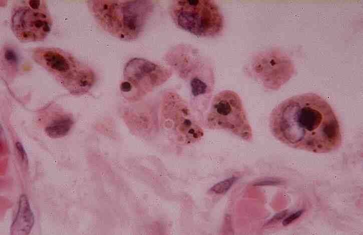
Lung: Chronic venous congesion
1.
Describe the key histopathological features shown in this slide.
Congested
pulmonary capillaries
An
intra-alveolar granular pink precipitate is seen.
In
acute pulmonary congestion, there may be associated alveolar
septal oedema or focal
intra-alveolar hemorrhage
In
chronic pulmonary congestion, the septa become thickened and fibrotic and
alveolar spaces may contain numerous hemosiderin-laden macrophages.
2.
Give a common cause for
pulmonary oedema.
Left-sided congestive heart failure
3.
How
would the lung function be affected?
Impairs the normal respiratory function.
Increases the thickness of the barrier between the blood capillaries and
alveolar spaces.
Predisposes lung to infection.
4.
What
is the macroscopic appearance of the lungs?
Hemorrhagic and wet
5.
List
5 pathophysiologic categories of oedema.
Increased
hydrostatic pressure
Reduced
plasma osmotic pressure
Lymphatic
obstruction
Sodium
retention
Inflammation
6.
In
congestive cardiac failure, which of the following statements are true?
There
is an absolute increase in plasma volume
T
There is an absolute increase in plasma Na content
F
There is an absolute increase in aldosterone secretion
T
There is a decrease in cardiac output. T
<< PREVIOUS INDEX NEXT SLIDE >>
Copyright � Joseph Ong 2003