Below is a collection of images designed to help you learn the anatomy and physiology of the horse. Because of the sheer size of some of these images, there was no way I could put them in full just on this page alone, so what you see below are just thumbnails. To view an enlarged version of an image, just click on it. Please feel free to print off any of these images to help you study equine anatomy. However, if you would like to use one of the below images on your website, please email me first to ask for permission – chances are I’ll let you use it but I just want to know where it’s being used at! J
Please keep in mind while scanning this information that this section of TTH has just begun – there may not be too much information contained here now but these pages will continue to grow into a very valuable resource. Anyway, happy studying!
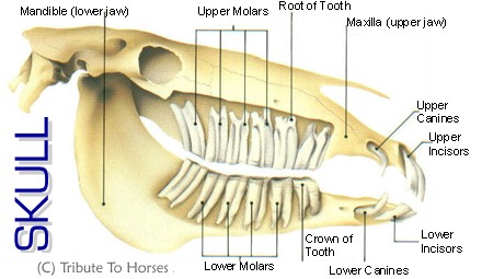
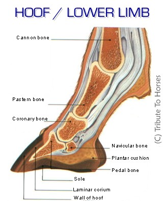
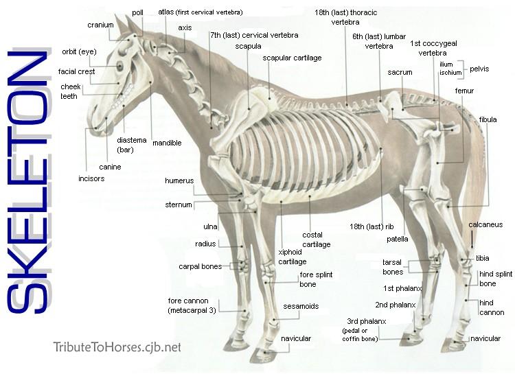
SKULL HOOF/LOWER LIMB * SKELETON
(this one may take a little longer to load… )
The bone structure of the horse's lower limb differs quite a bit from that of cats, dogs, and even humans. Cats, dogs, and humans all have several metacarpal(of the forelimb)/metatarsal(of thehindlimb) bones, while the horse has mainly only one - what we call the cannon bone is actually Metacarpal/Metatarsal III. The splint bones are vestiges of metacarpal/metatarsal bones, and are numbered as II & IV. Because of this single main metacarpal/metatarsal bone, the horse also only has one single digit (what we call a "finger") on each of his legs, made up of three phalanges - the proximal phalanx (long pastern bone), the middle phalanx (short pastern bone), and the distal phalanx (coffin or pedal bone). Also, the navicular bone we are all so familiar with is actually a sesamoid bone - the distal sesamoid to be more specific - that is embedded in the tendon to act as a "bearing" over the articular (joint) surface between the middle phalanx and distal phalanx.
Back to Equine Studies \/
http://www.geocities.com/lakersports/horsefacts/horsefacts.htm