 |
Cytometry
Volume 35 Issue 1 1999 Pages: 2-10 |
Abstract
Introduction Methods Results Discussion |
|
Cytometry
Volume 35 Issue 1 1999 Pages: 2-10 |
Abstract
Introduction Methods Results Discussion |
| Immunohistochemical Quantitation of Androgen Receptor Expression using Color Video Image Analysis |
| Desok Kim 1, Christopher W. Gregory 2, Gary J. Smith 3 4, James L. Mohler 1 3 4 * |
| 1Department of Surgery, Division of
Urology, The Laboratories for Reproductive Biology, University of North
Carolina, Chapel Hill, North Carolina
2Department of Pediatrics, University of North Carolina, Chapel Hill, North Carolina 3Department of Pathology, University of North Carolina, Chapel Hill, North Carolina 4UNC-Lineberger Comprehensive Cancer Center, University of North Carolina, Chapel Hill, North Carolina |
| Keywords |
| Abstract |
|
Background: |
| The immunostaining features of the androgen receptor (AR) have been studied in prostate cancer (CaP) to predict the outcome of androgen deprivation therapies. We have developed an automatic video color image analysis system for quantitation of AR expression in large samples of prostatic nuclei. |
|
Methods: |
| Essential criteria of immunostaining have been examined to establish a linear relationship between AR protein content and mean optical density (MOD) of the immunoperoxidase-substrate reaction product. Titration of monoclonal AR antibody, F39.4.1, and concentration and reaction time of substrate were optimized using color video image analysis. The methodology was tested twice. First, CWR22 human CaP xenograft specimens, harvested from testosterone (T)-stimulated, castrated and T-resupplemented mice, were immunostained to demonstrate the dependence of AR expression on serum androgen levels. Second, AR expression was measured in archived clinical specimens. |
|
Results: |
In CWR22 tumor-bearing mice castrated for 6 days, AR MOD decreased
to 57% of T-stimulated, intact mice. After 72 hrs of T treatment, AR MOD
returned to the level measured in T-stimulated, intact mice. Sixteen radical
prostatectomy specimens and 16 transurethral resection of prostate (TURP)
specimens were double-labeled with F39.4.1 and anti-cytokeratin MAb (13 E12)
specific for basal epithelial cells. Benign epithelial cells exhibited
lower AR MOD in prostatectomy compared to TURP specimens (P <
0.01). Differences in AR immunostaining intensity may have resulted from
differences in tissue fixation of whole organ versus small tissue specimens. E12)
specific for basal epithelial cells. Benign epithelial cells exhibited
lower AR MOD in prostatectomy compared to TURP specimens (P <
0.01). Differences in AR immunostaining intensity may have resulted from
differences in tissue fixation of whole organ versus small tissue specimens. |
|
Conclusions: |
| AR immunostaining can be quantitated accurately using optimized immunohistochemical criteria and video image analysis. Cytometry 35:2-10, 1999. 1999 Wiley-Liss, Inc. |
*Correspondence to James L. Mohler, UNC-Lineberger Cancer Center, CB# 7295, University of North Carolina, Chapel Hill, NC 27599-7295.
Funding Agency: National Institutes of Health; Grant Number:
AG11343 (to J.L.M.), CA64865 (to D.K., J.L.M., and G.J.S.), P30-HD-18968
(DNA and tissue culture cores)
Funding Agency: American Foundation for Urologic Disease (to
C.W.G.)
Funding Agency: Merck U.S. Human Health (to C.W.G.)
| Methods and materials |
Color
Video Image Processing
 |
 |
 |
 |
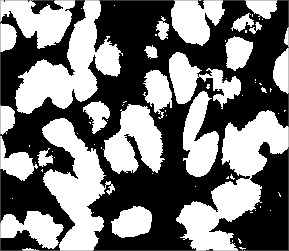
| Color image in RGB color scheme | Local adaptive thresholding [Ref. 23] and logical OR of RGB images | Classification of positive cells | Artifacts removed and mean optical density of cell nuclei assigned |
| References |
| 1 | Charalambous D, Kitchen PR, Stillwell RG, Smart PJ, Rode J. A comparison between radioligand and immunohistochemical assay of hormone receptors in primary breast cancer. Aust N Z J Surg 1993; 63: 637-641. Medline |
| 2 | Pertschuk LP, Schaeffer H, Feldman JG, Macchia RJ, Kim YD, Eisenberg K, Braithwaite LV, Axiotis CA, Prins G, Green GL. Immunostaining for prostate cancer androgen receptor in paraffin identifies a subset of men with a poor prognosis. Lab Invest 1995; 73: 302-305. Medline |
| 3 | Chang C, Kokontis J. Identification of a new member of the steroid receptor super-family by cloning and sequence analysis. Biochem Biophys Res Commun 1988; 55: 971-977. |
| 4 | Husmann DA, Wilson CM, McPhaul MJ, Tilley WD, Wilson JD. Antipeptide antibodies to two distinct regions of the androgen receptor localize the receptor protein to the nuclei of target cells in the rat and human prostate. Endocrinology 1990; 26: 2359-2368. |
| 5 | Lubahn DB, Joseph DR, Sar M, Tan J, Higgs HN, Larson RE, French FS, Wilson EM. The human androgen receptor: Complementary deoxyribonucleic acid cloning, sequence analysis and gene expression in prostate. Mol Endocrinol 1988; 2: 1265-1275. Medline |
| 6 | Zegers ND, Classen E, Neelen C, Mulder E, van Larr JH, Vorrhorst MM, Berrevoets CA, Brinkmann AO, van der Kwast ThH, de Winter JAR, Trapman J, Boersma WJA. Epitope prediction and confirmation for the human androgen receptor: Generation of monoclonal antibodies for multi-assay performance following the synthetic peptide strategy. Biochim Biophys Acta 1991; 1073: 23-32. Medline |
| 7 | Sar M. Application of avidin-biotin complex technique to the localization of estradiol receptor in target tissues using monoclonal antibodies. In: Bullock GR , Petrusz P , editors. Techniques in Immunocytochemistry, Volume 3. New York: Academic Press; 1985, p. 43-54. |
| 8 | Ruizeveld de Winter JA, Trapman J, Vermey M, Mulder E, Zegers ND, van der Kwast TH. Androgen receptor expression in human tissues: An immunohistochemical study. J Histochem Cytochem 1991; 39: 927-936. Medline |
| 9 | Magi-Galluzzi C, Xu X, Hlatky L, Hahnfeldt P, Kaplan I, Hsiao P, Chang C, Loda M. Heterogeneity of androgen receptor content in advanced prostate cancer. Mod Pathol 1997; 10: 839-845. Medline |
| 10 | Takeda H, Akakura K, Masai M, Akimoto S, Yatani R, Shimazaki J. Androgen receptor content of prostate carcinoma cells estimated by immunohistochemistry is related to prognosis of patients with stage D2 prostate carcinoma. Cancer 1996; 77: 934-940. Medline |
| 11 | Sadi MV, Barrack ER. Image analysis of androgen receptor immunostaining in metastatic prostate cancer. Heterogeneity as a predictor of response to hormonal therapy. Cancer 1993; 71: 2574-2580. Medline |
| 12 | Tilley WD, Lim-Tio SS, Horsfall DJ, Aspinall JO, Marshall VR, Skinner JM. Detection of discrete androgen receptor epitopes in prostate cancer by immunostaining: Measurement by color video image analysis. Cancer Res 1994; 54: 4096-4102. Medline |
| 13 | Prins GS, Sklarew RJ, Pertschuk LP. Image analysis of androgen receptor immunostaining in prostate cancer accurately predicts response to hormonal therapy. J Urol 1998; 159: 641-649. Medline |
| 14 | Benno RH, Tucker LW, Joh TH, Reis DJ. Quantitative immunocytochemistry of tyrosine hydroxylase in rat brain. I. Development of a computer assisted method using the peroxidase-antiperoxidase technique. Brain Res 1982; 246: 225-236. Medline |
| 15 | Nabors LB, Songu-Mize E, Mize RR. Quantitative immunocytochemistry using an image analyzer. II. Concentration standards for transmitter immunocytochemistry. J Neurosci Methods 1988; 26: 25-34. Medline |
| 16 | Pretlow TG, Wolman SR, Micale MA, Pelley RJ, Kursh ED, Resnick MI, Bodner DR, Jacobberger JW, Delmoro CM, Giaconia JM. Xenografts of primary human prostatic carcinoma. J Natl Cancer Inst 1993; 85: 394-398. Medline |
| 17 | Gregory CW, Sharief Y, Hamil KG, Hall SH, Pretlow TG, Mohler JL, French FS. Apoptosis in an androgen-dependent xenograft model derived from a primary human prostatic carcinoma [abstract]. Mol Biol Cell 1995; 6: 240. |
| 18 | Nagabhushan M, Miller CM, Pretlow TP, Giaconia JM, Edgehouse NL, Schwartz S, Kung HJ, de Vere White RW, Gumerlock PH, Resnick MI, Amini SB, Pretlow TG. CWR22: The first human prostate cancer xenograft with strongly androgen-dependent and relapsed strains both in vivo and in soft agar. Cancer Res 1996; 56: 3042-3046. Medline |
| 19 | Tan J-A, Sharief Y, Hamil KG, Gregory CW, Zang D-Y, Sar M, Gumerlock PH, deVere White RW, Pretlow TG, Harris SE, Wilson EM, Mohler JL, French FS. Dehydroepiandrosterone activates mutant androgen receptors expressed in the androgen dependent human prostate cancer xenograft CWR22 and LNCaP cells. Mol Endocrinol 1997; 11: 450-459. Medline |
| 20 | Wainstein MA, HE F, Robinson D, Kung H-J, Schwartz S, Giacona JM, Edgehouse NL, Pretlow TP, Bodner DR, Kursh ED, Resnick MI, Seftel A, Pretlow TG. CWR22: Androgen-dependent xenograft model derived from a primary human prostatic carcinoma. Cancer Res 1994; 54: 6049-6052. Medline |
| 21 | Brigati DJ, Budgeon LR, Unger ER, Koebler D, Cuomo C, Kennedy T, Perdomo JM. Immunocytochemistry is automated: Development of a robotic workstation based upon the capillary action principle. J Histotechnol 1988; 11: 165-183. |
| 22 | Mize RR, Holdefer RN, Nabors LB. Quantitative immunocytochemistry using an image analyzer. I. Hardware evaluation, image processing, and data analysis. J Neurosci Methods 1988; 26: 1-24. Medline |
| 23 | Otsu N. A threshold selection method from gray-level histograms. IEEE Trans Systems Man Cybernetics 1979; 9: 62-66. |
| 24 | Serra J. Image analysis and mathematical morphology. Boston: Academic Press; 1982. |
| 25 | Kim D, Charlton JD, Coggins JM, Mohler JL. Semi-automated nuclear shape analysis of prostatic carcinoma and benign prostatic hyperplasia. Anal Quant Cytol Histol 1994; 16: 400-415. Medline |
| 26 | Gonzalez RC, Woods RE. Digital image processing. Reading, MA: Addison-Wesley; 1992. p. 229-237. |
| 27 | Gregory CW, Hamil KG, Kim D, Hall SH, Mohler JL, French FS. Androgen receptor expression in androgen-independent prostate cancer is associated with increased expression of androgen-regulated genes. Cancer Res 1998, in press. |
| 28 | Bonkhoff H, Remberger K. Widespread distribution of nuclear androgen receptors in the basal cell layer of the normal and hyperplastic human prostate. Virch Arch A Pathol Anat Histopathol 1993; 422: 35-38. |
| 29 | Hobisch A, Culig Z, Radmayr C, Bartsch G, Klocker H, Hittmair A. Distant metastases from prostatic carcinoma express androgen receptor protein. Cancer Res 1995; 55: 3068-3072. Medline |