*Ado Oculus Imaging Classes
Personal
Links
Curriculum
Vitae
Chapel
Hill, NC
Research
Interests
Computerized
cytometry
Watershed
transform
AdoIC
imaging library
CCD
Camera
Seminar
If you have comments or suggestions, email me at [email protected]
![]()
This page was created with Netscape Navigator Gold
This counter service is provided by Ultimatecounter.com
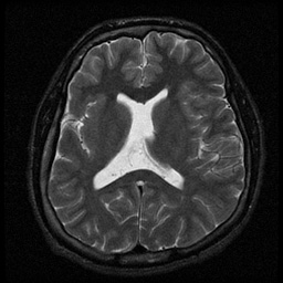
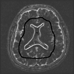
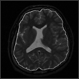
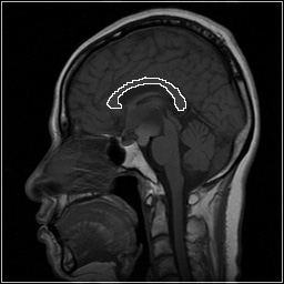
This page illustrates AdoIC's interactive watershed segmentation that has been suggested by F. Meyer.
Interactive Meyer's Watershed
 |
 |
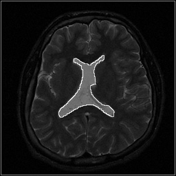 |
| The axial MRI image of the brain (top left) was dilated or eroded (not shown). Their difference was scaled up from 50 to 255 and markers (gray level=0) were imposed interactively (top middle). Meyer's watershed found the outline of the lateral ventricle (top right). Markers at the lower center image were imposed to find the outline of the brain gray matter (lower left). |  |
 |
 |
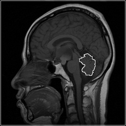 |
The corpus callosum (left) and the cerebellum (right) were some of distinctive features in the sagittal image of the brain. In this type of the brain image, the whole brain is not well outlined. Meyer's watershed does not detect the ideal contour of the brain mainly due to the strong gradient between the scalp and the skull. |