The Medical Radiation course leads to three career pathways. In the 2nd semester of the 1st year the medical radiation separates into different streams, which includes the Medical Imaging stream, Nuclear Medicine stream and the Radiation Therapy stream.
I have finished my undergraduate degree and I am currently working as an Intern.I hope to continue with further studies / post graduate studies in Ultrasound after this achievement. This involves the learner to be working in a full time department as an ultrasonographer.
Well lets see what is in store for me as life takes me.........
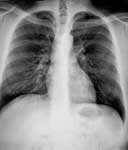
Radiography is a vital health profession. It assists in the diagnosis and management of human illness by producing images of the structures in the body. Many people are quite familiar with a radiograph or an "x-ray". You may well have had such an examination yourself. Unlike photography which uses light waves in its work, radiography has traditionally used that part of the electromagnetic spectrum known as x-rays to produce a radiograph such as this showing the chest.
With the use of special contrast agents in conjunction with x-rays, radiographs of many of the organs of the body can be produced such as this image of the kidneys.
As well as producing images of the body, these modalities are also used in conjunction with additional equipment, to identify and treat a variety of disorders such as narrowing of the blood vessels.
What do Radiographers do?
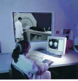
Radiographers are health professionals who have the knowledge and understanding required to use and manipulate radiographic equipment and complex medical imaging technology to generate a variety of images for subsequent interpretation and storage.
Generally speaking they work as part of a medical imaging team. Using x-ray film-based imaging, radiographers select and implement the most appropriate examination protocol which will deliver the lowest possible dose of radiation to the patient.
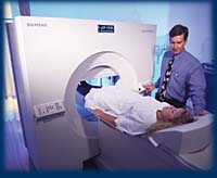
Whilst a CT scan still uses x-rays, unlike the familiar radiograph, CT does not use photographic screens and film to detect the attenuated x-ray beam as it leaves the patient. Instead this radiation is collected by specialised detectors and transmitted to a computer for processing.
The image software capabilities of CT generate cross-sectional images.
Ultrasound uses high frequency sound waves and a specialised probe to generate images which are different from an x-ray.
Because of the technical nature of the medical imaging environment, patients look to radiographers for advice and assurance throughout their examination.
In order to perform their professional role, radiographers must therefore be competent in radiographic and medical imaging science and methods, radiologic physics, radiation protection, and radiologic biology. They must also be able to care for the patients undergoing radiographic and medical imaging examinations.
The movement of organs and blood vessels can be observed directly on a television monitor and the body can be imaged in any plane. A form of ultrasound known as Doppler imaging can allow the velocity of blood to be estimated at different sites within the two-dimensional image and measurements to be calculated.
General radiography involves assessing the patient history, making a clinical diagnosis and taking appropriate radiographs to demonstrate the relevant anatomy. The production of radiographs, it is not simply pressing a button. An understanding of the interaction of x-rays with matter and an accurate precise delivery of radiation has to be assessed in order to gain an image of diagnostic quality.
Emergency radiography requires an adaptation of general radiographic skills and putting them into action into the trauma/resuscitation scene of the emergency department. Trauma patients presenting to the emergency department are often traumatised or debilitated. Their presentation may be the result of acute or chronic injury or condition as the result of a car accident, heart attack, sport injuries or general sickness. As a trauma radiographer, you have to be able to cope or learn to cope with some very unsightly graphic and disturbing sights and emotions. You have to cope with death, gruesome sights of blood and body disfigurement, burns victims and an often-heady pace as an integral member of the health professional team. Emergency radiography technically differs for the radiographer because the patients often are not allowed to move at all, which means you have to get the same images required of the anatomy without moving the patient.
Computed Radiography scanning, uses x-radiation to gain cross sectional images of human anatomy. C.T. is an incredibly sensitive modality that gives us a three-dimensional view of virtually all tissue types, organs and visceral boundaries.
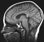
M.R.I. unlike C.T. uses magnetic fields and radio frequency to image or reconstruct anatomy in any plane. M.R.I. is even more sensitive than C.T. for most tissue types. Nerves, muscles, cartilage and even bodily fluids can be detected without the need for injection to aid diagnosis.
M.R.I. works by aligning the atoms of the body to an external magnetic field of intensity between 1 and 2 Tesla. An external radiofrequency is beamed into the tissue of interest. This causes the atoms to precess (spin) around a north axis. When the radiofrequency pulse is removed the precessing slows until they relax back into the north aligned position. Each different tissue relaxes at different rates, since there is a changing magnetic field inside the patient this is picked up by external coil detectors as an induced current. Thus all the different tissues may be differentiated by the amount of precession. M.R.I. is starting to eliminate a lot of painful, interventional procedures.
Fluoroscopic X-Ray images the gastro-intestinal system and specialised bodies systems. The advantage of Fluoroscopic x-ray over conventional x-ray is that you can view in real time on a television monitor the image. This means that movement of the peristalsis and normal function of the body's systems can be captured and recorded on videotape or videodisc. Procedures that are most commonly done with the aid of fluoroscopy include barium enemas, barium meals, myelograms (spinal cord imaging) and fracture reduction.
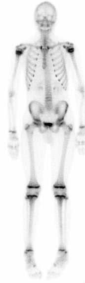
As an integral part of patient care, nuclear medicine is used in the diagnosis, management, treatment, and prevention of serious disease. Nuclear medicine imaging procedures often identify abnormalities very early in the progression of a disease -long before some medical problems are apparent with other diagnostic tests. This early detection allows a disease to be treated early in its course when there may be a more successful prognosis.
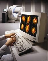
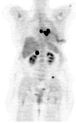
Nuclear medicine uses very small amounts of radioactive materials or radiopharmaceuticals to diagnose and and treat disease. Radiopharmaceuticals are substances that are attracted to specific organs, bones, or tissues. The radiopharmaceuticals used in nuclear medicine emit gamma rays that can be detected externally by special types of cameras: gamma or PET cameras. These cameras work in conjunction with computers used to form images that provide data and information about the area of body being imaged. The amount of radiation from a nuclear medicine procedure is comparable to that received during a diagnostic x-ray.
Today, nuclear medicine offers procedures that are helpful to a broad span of medical specialties, from pediatrics to cardiology to psychiatry. There are nearly one hundred different nuclear medicine imaging procedures available and not a major organ system which is not imaged by nuclear medicine.
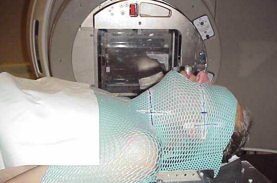
Only the cells in the area that is being treated are affected by the radiation. Although both normal and diseased cells are damaged, normal cells have the ability to regenerate and repair, unlike the diseased cells which can not repair themselves. As the treatment progresses more of the diseased cells die and are not regenerated causing the mass to shrink, whereas the normal cells are being continually replaced as part of normal body function. The cells that die are broken down and excreted by normal processes of the body.
The aim of Radiotherapy treatment is to destroy diseased cells while limiting the impact on normal cells and thereby decreasing the side effects to a manageable level. This is achieved by delivering the maximum amount of dose to the diseased cells and minimising the dose to normal tissue. It is the job of the Radiation Therapist to design specialised treatments that may involve shielding normal tissue, various treatment angles, and individualised stabilising devices inorder to attain this aim.