 |
| Figure 1 (left) shows the structure of a complex formed by the platelet receptor GpIb(alpha) and thrombin that we recently determined. Direct interaction between GpIb and thrombin is required for platelet aggregation and activation at sites of vascular injury. Abnormal GpIb-thrombin binding is associated with many pathological conditions, including occlusive arterial thrombosis and bleeding disorders. The crystal structure presented above reveals simultaneous interactions of GpIb with exosite I of one thrombin molecule and with exosite II of a second thrombin molecule. In the crystal lattice, the periodic arrangement of GpIb-thrombin complexes mirrors a scaffold that could serve as a driving force for tight platelet adhesion. The details of these interactions highlight two distinct interfaces that are potential targets for development of novel antithrombotic drugs. This work was published in Science (summary; reference). |
| This site features highlights of my recent postdoctoral work. I am a Research Scientist interested in determining the three-dimensional structures of proteins and protein complexes that are targets for structure-based drug discovery using the powerful technique of X-ray crystallography. I currently work at Wyeth (formerly Genetics Institute) in Cambridge, MA. More information can be found on my resume.... |
| Figure 3 (right) shows the overall structure of the homodimeric C-terminal region of Early Endosome Antigen 1 (EEA1) bound to Ins(1,3)P2, with the polypeptide chains colored green (chain A) or blue (chain B), the head group colored yellow (carbon and phosphorus atoms) and red (oxygen atoms). The molecular surface is rendered semi-transparent and corresponds to the individual polypeptide chains. The overall structural features of EEA1 include an extensive coiled-coil, a calmodulin-binding motif, and two Zn++binding motifs, one at the amino terminus and the other at the carboxyl terminus (Fig. 1). EEA1 is required for endosome fusion. Ins(1,3)P2 is the soluble portion of a phospholipid found in lipid membranes. This structure lets us better understand precisely how EEA1 interacts with the surface of endosomal membranes. This work was published in Molecular Cell (reference). |
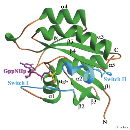
| Figure 4 (right) shows the structure of the active form of the GTPase Rab3A, a protein which regulates exocytosis and neurotransmitter release. This work was published in Structure (reference). |
 |
| John J. Dumas, Ph.D. |
| Home |
| Miscellaneous Links |
| Figure 3: The Crystal Structure of Ins(1,3)P2-bound EEA1 |
| Figure 4: The Crystal Structure of GTP-bound Rab3a |
 |
| Figure 1: The Crystal Structure of the GlycoproteinIb(alpha)-thrombin Complex |
| (Figures 3 & 4 depict structures that were determined when I worked at UMass Medical School in the Program in Molecular Medicine.) |
| Since 1993 I have lived in Arlington, MA. A historic treasure located between Cambridge and Lexington, Arlington is the birthplace of "Uncle Sam" and is home to a vibrant, culturally diverse community. Favorite sites of interest include the Capitol and Regent Theaters, the Minuteman Bike Trail, and many great restaurants along "restaurant row" on Mass. Ave. |
 |
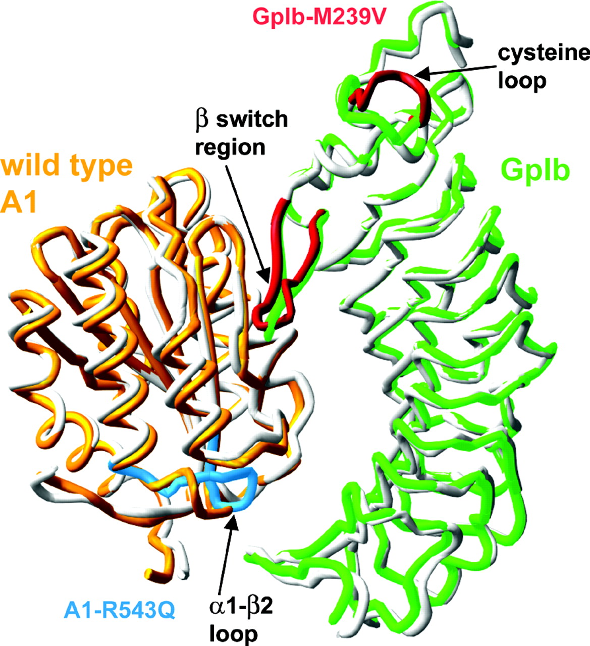 |
| Figure 2 (above) The adhesion of blood platelets at the site of vascular injury under high shear stress is mediated by a direct interaction between the platelet receptor GpIb(alpha) and the A1 domain of the blood glycoprotein von Willebrand factor. Direct comparison of the wild-type GpIb-A1 structure to that of a complex comprised of proteins containing two Von Willebrand Disease mutations reveals specific structural differences between these complexes at sites near the two binding interfaces. These findings provide important details that add to our understanding of how these mutations affect binding affinity in this common bleeding disorder. This work was recently published in the Journal of Biological Chemistry (reference). |
| Figure 2: The Crystal Structure of the wild-type GpIb-A1 complex |