Dynamic Anatomy by Burne
Hogarth
This is a great book to use for modeling. Hogarth breaks down the human body into a simple system of planes. It makes modeling a human much more understandable. You should pick up the book, but you don’t have to. Here are some images that might help you as you work on your human head.
THE EYE
|
|
|
|
|
The axis of the eye. The upper lid arches high while the lower lid curves to the outside of the socket. |
The eyelids appear as visors surrounding the orb. |
Jenga’s note: Notice the thickness of the lid here and
the sharp definition of the outer and inner edge. |
|
|
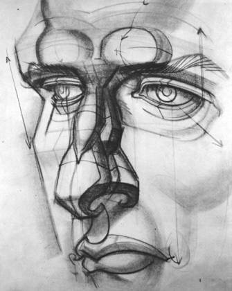
Left: The eye position, side view, starts on a line drawn up from the nostril wing. Right: The axis of the eye. The upper lid arches high while the lower lid curves to the outside of the socket. |
|
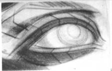
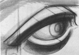
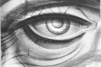
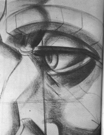
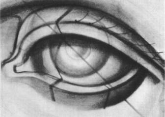
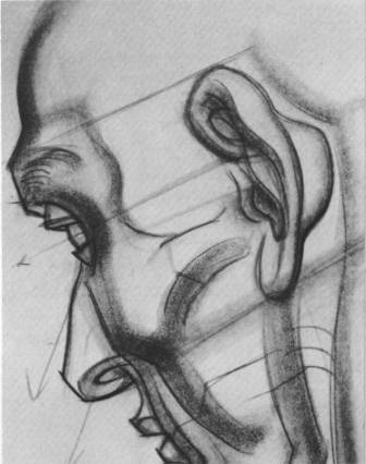
The Eye: The eyeball, almost as large as a golf ball, in the human head, is a n exposed internal organ of the body protected by great structures of bone, the brow ridge (superciliary arch) and the cheek bone (zygomatic bone). It is suspended from the roof of the eye socket (orbit). The eyelids curve like short visors on the eye; the upper curves wider across the fuller circumference of the orb, while the lower turns on a shorter arc, around the base area. Seen from a side view, the lower lid lies angled down almost 45 degrees from the upper lid. Surrounding the eye is the orbicularis muscle, enclosing and circling the orbit. It gives little shape, however, to the surface form of the socket and cheek bone.
THE NOSE
|
|
The Nose: The nose consists of four important masses: the upper nasal mass, tapered and wedged into the cartilaginous (alar) ball of the nose, and the two wings (ala) of the nostrils. The ball of the nose swings into a furrowed hook (septum) under the base and meets the pillars of the upper lip. THe nostril wings, moving from the ball of the nose, flare out to the sides, the length of an eye apart. The nostril cavities are triangulated in shape and should be drawn large enough to accommodate the thickness of a finger. |
|
|
|
|
|
THE LIPS
|
|
|
|
|
|
|
|
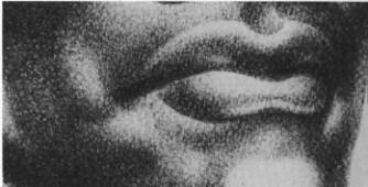
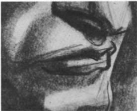
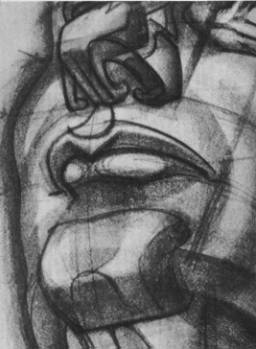
The Lips: The lips are surrounded by a sphincter muscle (orbicularis oris) and attach at the sides of the mouth to the buccinator muscle, which crosses horizontally from the jaw. The upper lip, wider than the lower, is shaped like a flattened “M.” The groove of the “M” (philtrum) thrusts forward like the prow of a ship (tubercle). The lower lip is developed like an extended “W.” The center groove receives the tubercle of the upper lip, while the arms of the “W” form two elliptical lobes. Both lips have thinly-edged margins which rim their forms.
THE EAR
|
|
|
|
|
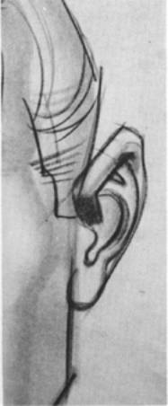
Jenga’s note: Notice the way the ear is divided into
planes. This looks like a poly model
before it’s converted to Sub-D, doesn’t it?
Think about this when you’re modeling. You’re creating distinct planes. |
|
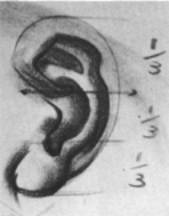
The ear divides into equal thirds lengthwise. |
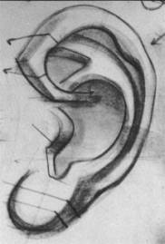
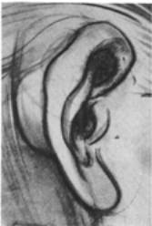
The Ear: The ear is shaped like a shell, wider at the top
rim, narrower at the lobe. It consists
of four major shapes: the outer wider
rim (helix), the inner rim (antihelix), the cover of theear opening (tragus),
and the lobe (lobule). The ear can be
divided into equal thirds lengthwise: first, at the upper rim where it enters
the bowl of the ear; second, the length of the tragus; third, the fleshy
lobe. The inner rim is divided at the
top into two arms and shaped like a bent “Y.”
Emphasis in drawing should be given to the hard forms of the cartilage,
and softened on the fleshy lobe. The
bowl of the ear should be drawn large enough to accommodate a thumb.
|
|
|
|
|
|
|
|
Updated June 10, 2002 by Jenga Mwendo