
Go to the
linked sites to know more about the topic If you have
an outstanding image of philatelic interest send it to me by e-mail, and I will publish it here.
 Stamp of the month
| These stamps honors Dr. George N. Papanicolaou, 1883-1962, the father of cervical cancer screening by cytology, the Pap test. The USA one was issued to commemorate the 100th anniversary of his birthday. Images sent by Dr V. Schneider, courtesy of Dr. Bernard Naylor. Visit this site to see an outstanding collection of stamps depicting microscopes | 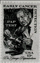 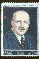 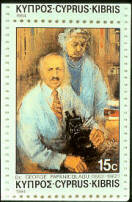 USA, 1983 - Greece, 1973 - Cyprus, 1984 |
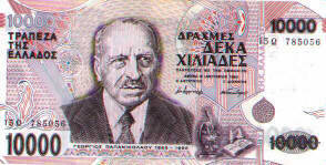
| Numismatic item: This is a 10,000 dracma bill of Greece honoring Dr. Papanicolaou Image sent by Volker Schneider, Germany |
|
..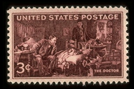
| This stamp features "The Doctor", the famous painting by Samuel L. Fildes. It was issued to commemorate the 100th anniversary of the founding of the American Medical Association.
USA, 1947 Link: Moody Medical Library |
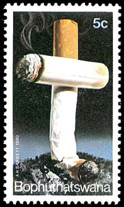 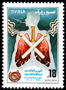 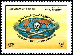 |
Stamps on Fight against smoking
|
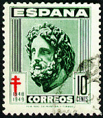
ASCLEPIOS, Spain 1948 Link: http://www.webcom.com/mjljweb/jrnlclb/stamps/asclepios.html
| 
|
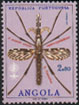 .. ..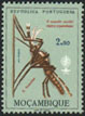  |
Stamps on Malaria eradication
Angola, 1962 - Mozambique, 1962 - Link: http://www.malariastamps.com/
|
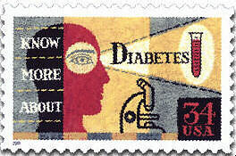  |
USA, 2001 Diabetes awareness, |
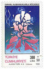 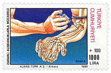 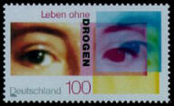 |
Stamps on Fight against drugs
Turkey, 1990 Two stamps |
 | Stamp honoring Dr Virginia Apgar,
of APGAR score fame
USA, 1994 |
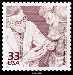 |
Stamp honoring Dr Jonas Salk, of polio vaccine fame |
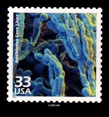
..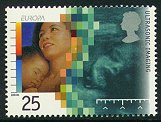 .. 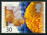 .. 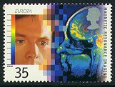 .. 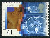 | Ultrasonic Imaging - 25p -ultrasound scans for pregnant women are now a standard test throughout much of the world. Sonar technology, first used to find submarines in the 2 world wars, was turned to this medical use by Prof. Ian Donald, at Glasgow Univ. Scanning Electron Microscopy- 30p -these microscopes take images at a scale of less then a few thousandths of a mm across. One use is to examine blood cells for abnormalites, such as found in sickle-cell anemia. The machines are a product of early research at Cambridge Univ. under Charles Oatley. Magnetic Resonance Imaging - 35p -this process makes use of the fact that the human body is comprised of mostly water, that diseased and healthy tissue have different amounts of water and that cancer tissue produces its own MRI trace. The process is a result of work at Aberdeen and Nottingham Universities by Profs John Mallard, Peter Mansfield and Raymond Andrew. Computed Axial Tomography - 41p -also known as the CAT scan, uses precise beams of x-rays to take pictures of a slice, or one particular plane, of the body. A computer puts together a picture of the area scanned. Godfrey Hounsfield shared the Nobel Prize for Medicine in 1979 for his work with the CT. UK, 2000 - The stamps shown here were issued by the British Royal Mail to recognize the part United Kingdom scientists played in these advances. |
 ......
 ..
..