Adenocarcinoma, papillary, metastatic in pericardium:


Drs. Prolla and Diehl's INTERESTING CASE OF THE MONTH December 2001 case B

Adenocarcinoma 100x Pap staining, acinar and papillary groups of cells, and typical psammoma bodies

200x Pap staining

200x Pap staining, the insert shows typical psammoma bodies

Adenocarcinoma 500x Pap staining


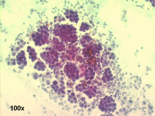
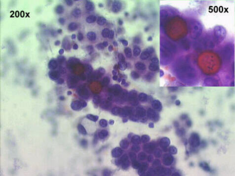
Adenocarcinoma, papillary, Pericardial fluid, cardiac tamponade in 66-year old male

The smears show a typical papillary carcinoma, with massive number of acinar and papillary groups of cells, and many psammoma bodies. The patient had a bronchogenic carcinoma, with lymphangitic spread and pericardial metastases. The signs of cardiac tamponade (narrowed pulse pressure, distended neck veins, muffled heart sounds, and pulsus paradoxus) were relieved by the pericardiocentesis. One week later, a pericardial window procedure was done.


Features of Papillary Adenocarcinoma

Cellular patterns:
Three-dimensional groups, papillary arrangement, acinar, cell balls
Presence of psammoma bodies
Few single cells
Cytoplasm:
Basophilic, finely or coarsely vacuolated, cuboidal or round cells
Eccentrically placed nuclei, touching the cellular borders
Nucleus
Irregular chromatin pattern, with some coarse chromocenters
Prominent single or multiple nucleoli


See also Case A

References
List of cases
Atlas
Hosted by www.Geocities.ws

1