Drs. Prolla and Diehl's
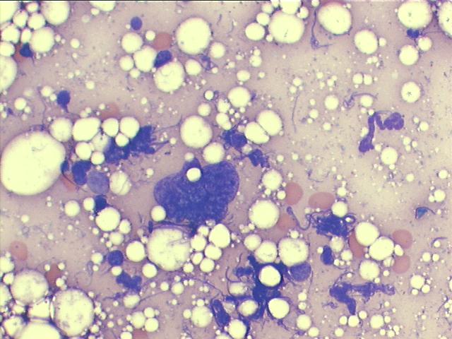
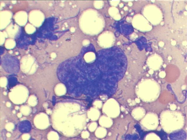
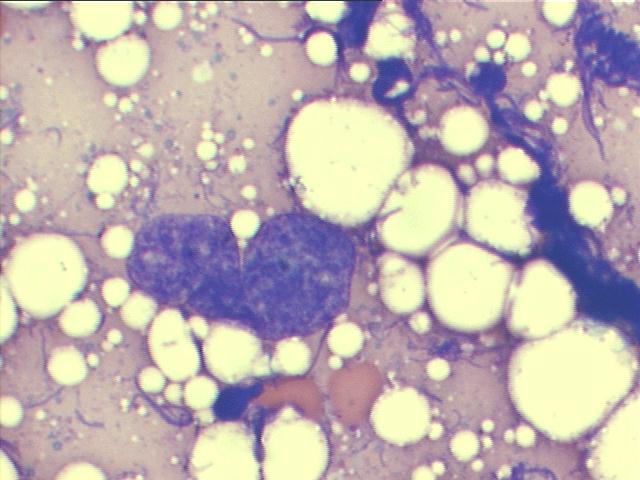
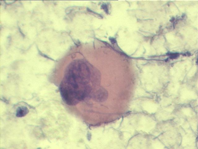
INTERESTING CASE OF THE MONTH December 2007 FNA of paravertebral mass: thoracic extramedullary hematopoiesis
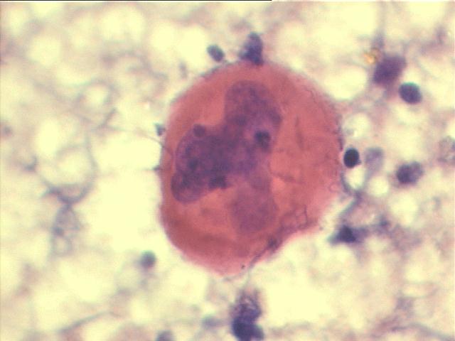
The smears show a great number of megakaryocytes, in a background of hematopoietic cells and fat. The computed tomography of the chest demonstrated soft tissue density masses in the lower third of the paravertebral spaces. These masses had well-defined limits, lobulated edges and were heterogeneous due to areas of interposed soft density tissues and adipose tissue, considered typical of extramedullary intrathoracic hematopoiesis. The use of needle biopsy in this condition is controversial due to the risk of hemorrhage (Dal Pozzo G, Moroni F, Pellegrini GL, Villari N. Computed tomography role in massive thoracic extramedullary haematopoiesis. Eur J Radiol 1982; 2:235-7.) This is a rare case of intrathoracic extramedullary hematopoiesis documented by FNA. The fine needle technique was considered safe by us.