The mesenteric node imprint showed several larval forms of Strongyloides stercoralis, a quite unusual finding, never reported before in cytological specimens. Photos 1 and 2 show the presence of many Strongyloides stercoralis larvae in the mesenteric node specimen histology. Photos 3 and 4 show massive small bowel infiltration by the same larvae. Hyperinfection syndrome by Strongyloides stercoralis had been reported in AIDS patients before, but the cytological findings are here reported by the first time.
Hospital de Clinicas de Porto Alegre, Cytopathology Laboratory
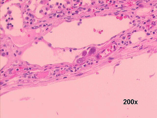
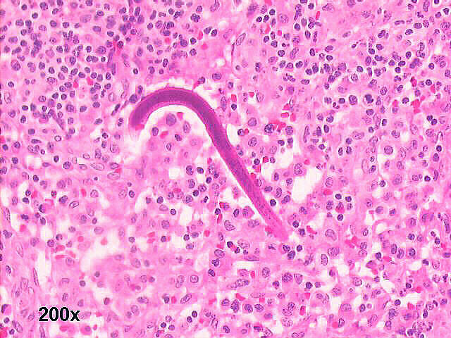
Photos 1 and 2: Lymph node histology, H&E staining (see above)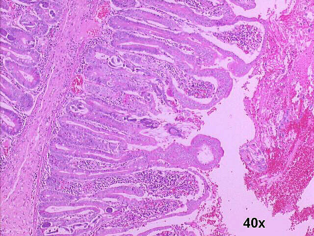
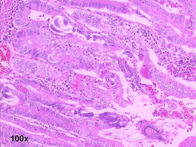
Photos 3 and 4: Small bowel resection specimen histology, H&E staining(see above)
Porto Alegre,
RS Brazil