 ascites
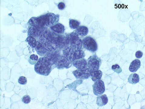 ascites
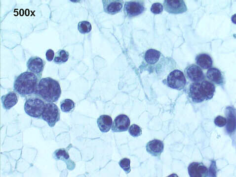 ascites
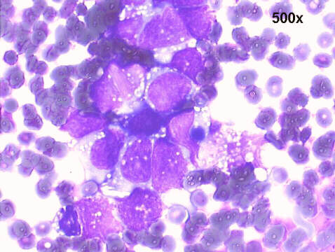 ascites
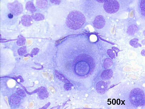 groin mass
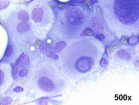 groin mass
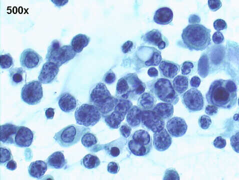
The ascitic fluid had many loose aggregates of small malignant cells, undifferentiated pattern, with coarse chromatin, and in the M-G-G staining had small cytoplasmic vacuoles. The mass in the left groin had much larger malignant cells, many of them with intranuclear vacuoles, quite suggestive of amelanotic malignant melanoma metastases. A surgical biopsy of a large retroperitoneal mass was interpreted as metastases of amelanotic malignant melanoma, and its cellular pattern was quite similar to the ascitic fluid sediment, and at immunohistochemistry was positive for S-100, and also for HMB 45. This case is remarkable for the small cell malignant melanoma pattern in the ascitic fluid and the more classical large cell pattern in the groin mass.

| Case A Oct 2008 | References |
|
Atlas |