|
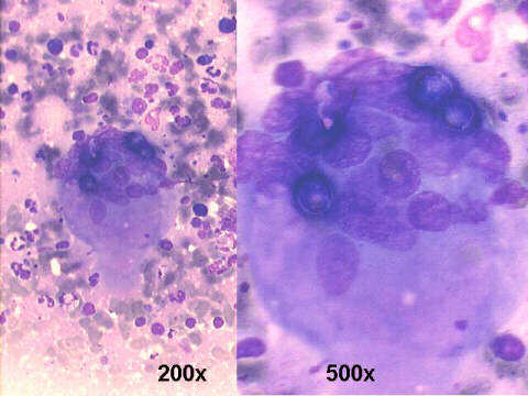
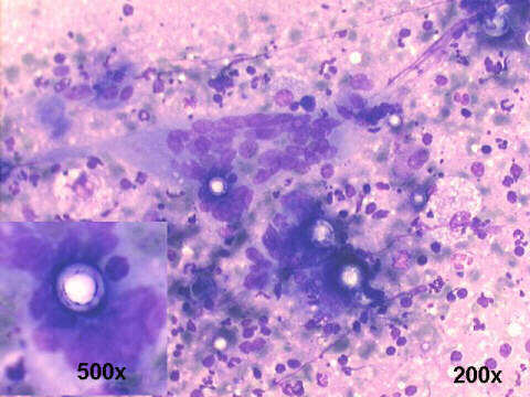
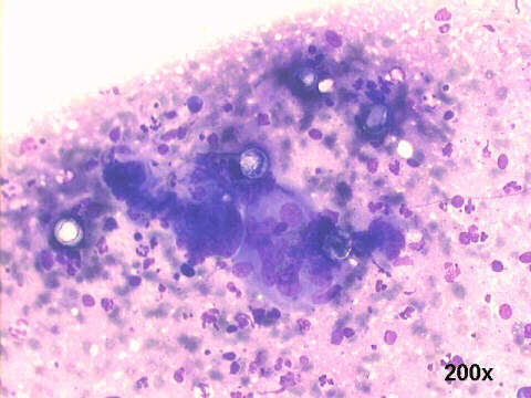
The M-G-G stained smears show many giant cells which contain the round organisms with refractile walls. The fungus occurs as a budding yeast with cells 18-24 um in diameter, but some are quite large (over 40-60 um), and many extracellular fungi are present. The Gomori staining of the surgical biopsy and the cultures confirmed the diagnosis. Ulcerated oral lesions are frequent, as well as enlarged neck lymph nodes, in the advanced systemic infection stage of the disease. But, the disease is always acquired by inhalation of the microorganism.
|