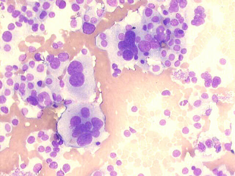
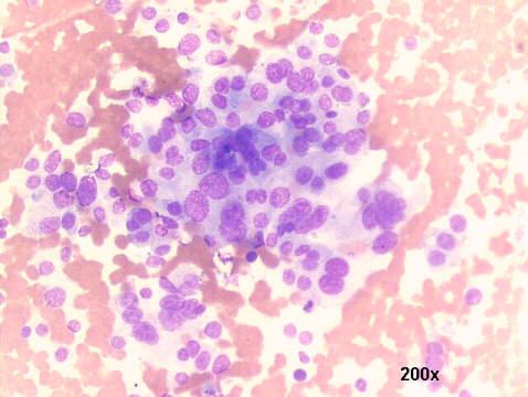
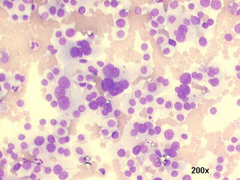
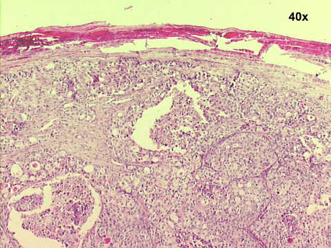
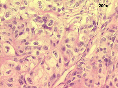
Cytology features: smears very cellular, with absent colloid. The follicular cells occur in sheets, but mostly as dispersed single cells, with little atypia, except for marked anysonucleosis. Histology revealed a follicular carcinoma of the thyroid, with a trabecullar pattern. The 40x image shows the tumor capsule, while the 200x image shows the cellular atypia. (H&E staining)