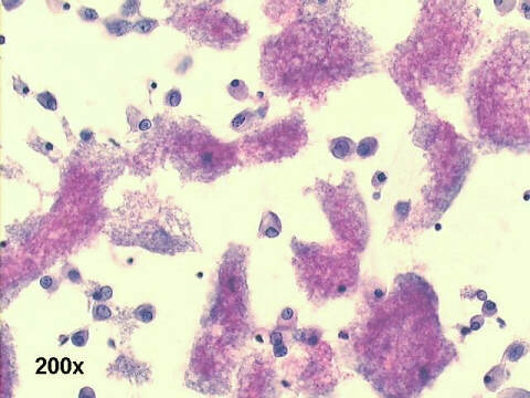
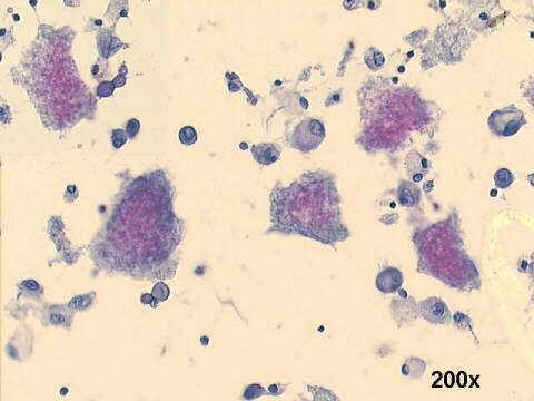
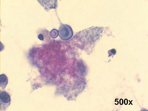
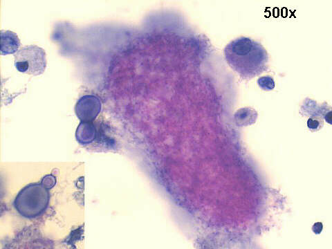
Broncho-alveolar lavage, 37-year old man with AIDS
Double infection: Pneumocystis jiroveci and Cryptococcus neoformans
>
This case shows two organisms highly prevalent in lung infections of AIDS patients.
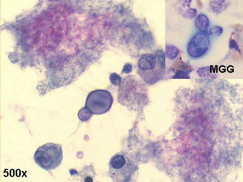
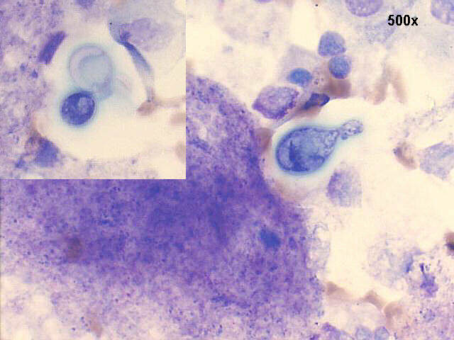
1)Cryptococcus. The fungus is spherical, and produces a single teardrop shaped bud. The thick mucoid capsule produces an unstained halo around the organism, in Papanicolaou stained smears. In M-G-G stained smears the capsule may stain in pink color, due to its high sugar content, and the fungi are more easily seen, even if in low numbers.
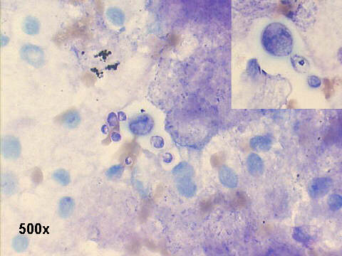
2)Pneumocystis. The Papanicolaou or M-G-G stained material showed the typical alveolar casts, with a foamy appearance, highly suggestive of P. jiroveci pneumonia. The name of this fungus has been recently changed to Pneumocystis jiroveci, and the former name P. carinii is now used only for the organisms found in rats.
