|
|
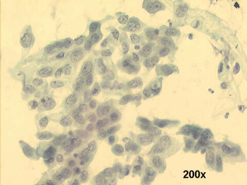
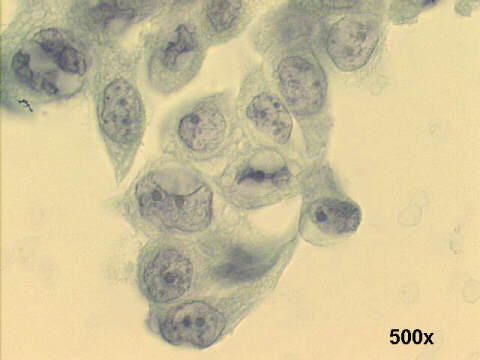
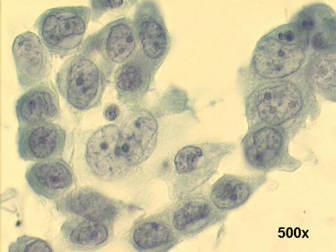
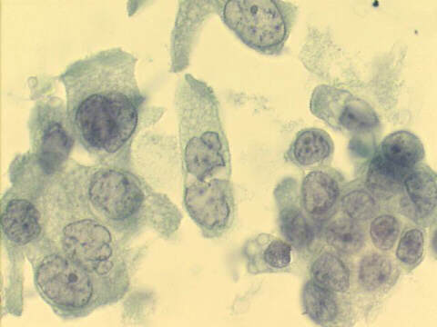
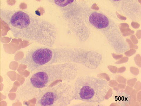
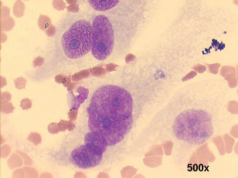
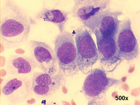
The patient had typical Barrett's esophagus at endoscopy, with circular involvement, over 4cm in length. The smears show abundant number of columnar cells, with abnormal looking nuclei (nuclear enlargement with abnormal shapes, conspicuous nucleoli, chromatin irregularity) and abnormal cytoplasm: loss of mucin production, and cellular dyscohesiveness, suggestive of high grade dysplasia. This was confirmed by endoscopic biopsy, and also after surgical biopsies. The patient is on endoscopic surveillance, after endoscopic ablation of his lesion, and an anti-reflux operation.

| Case B September 2003 | Case C September 2003 | References | List of cases | Atlas |