Drs. Prolla and Diehl's CASE OF THE MONTH August 2007
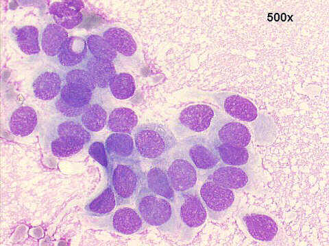
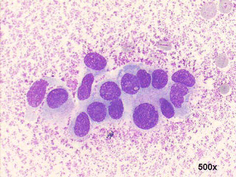
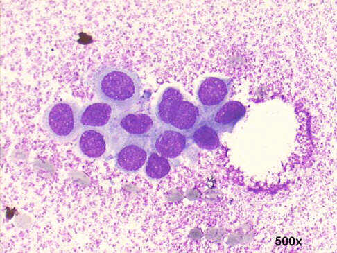
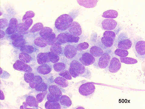
FNA breast nodule, 66-year old male: gynecomastia (false positive cytology)
Drs. Prolla and Diehl's CASE OF THE MONTH August 2007FNA breast nodule, 66-year old male: gynecomastia (false positive cytology)
|
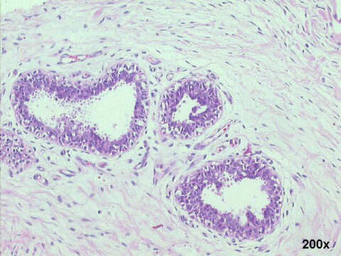
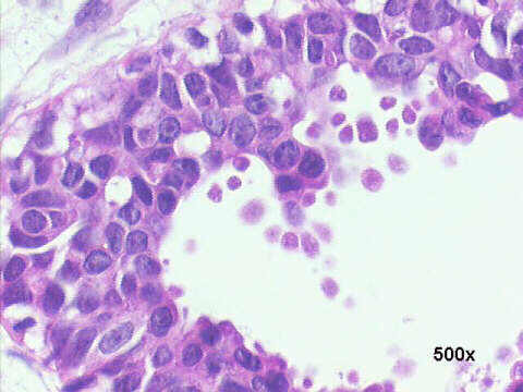
The smears show sheets of epithelial cells, no stroma nor myoepithelial cells. The evenly distributed chromatin, the absence of nucleoli, the regular oval shape of the nuclei, are noteworthy. However, the somewhat dyscohesive nature of the sheets, their relatively small size, and the absence of myoepithelial cells, made us to call the case a well differentiated ductal cell carcinoma. Surgery revealed the nodule to be gynecomastia with moderate atypical proliferation of the duct epithelium. One year later, the patient had a colonic carcinoma diagnosed and resected.

| Case August 2007 | References | List of cases | Atlas |