|
|
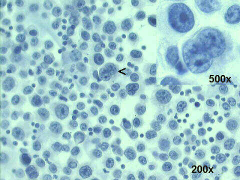
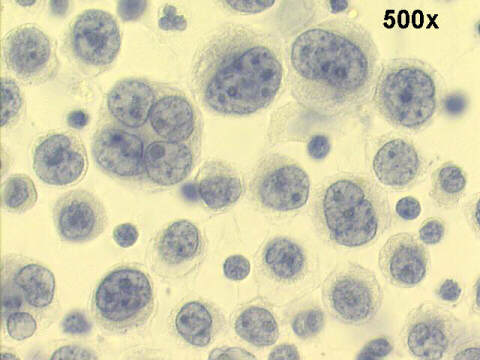
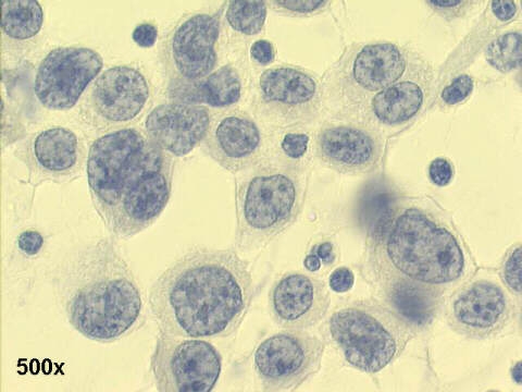
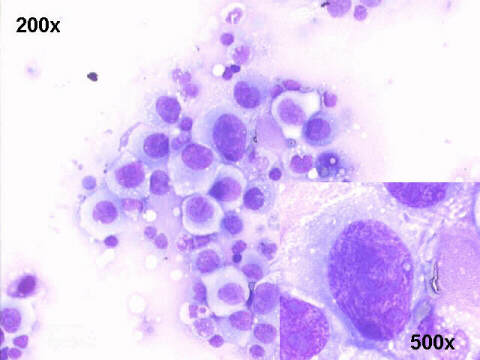
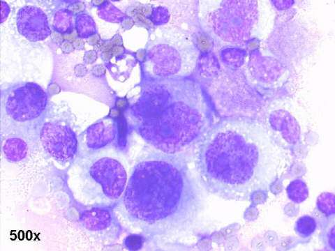
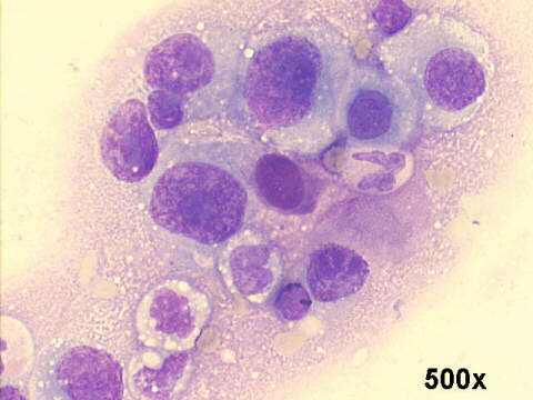
The smears show the typical features of metastatic undifferentiated carcinoma in ascites fluid sediment: many undifferentiated malignant cells, with variable size, large nucleoli and clumped chromatin. A few loose groups and sheets showing nuclear abnormalities, with variable amounts of cytoplasm. The patient had an inoperable stomach cancer.

| Case B August 2003 | Case C August 2003 | References | List of cases | Atlas |