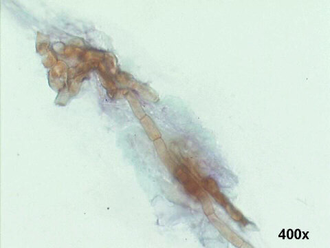
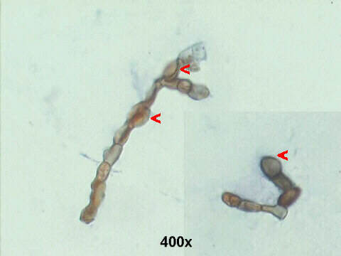
Presence of septated hyalin hyphae, with several chlamydoconidia, see the red arrow heads on the lower illustration. The fungus was seen in several repeated corneal samplings, and on culture was identified as Fusarium spp. It grew rapidly on Sabouraud dextrose agar at 25°C and produced cottony, flat, spreading colonies. Fusarium spp. is the commonest cause of opportunistic mycotic keratitis.
