Drs. Prolla and Diehl's Case of the month ~ July 2007 case A

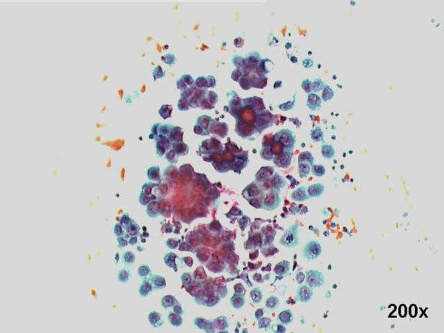
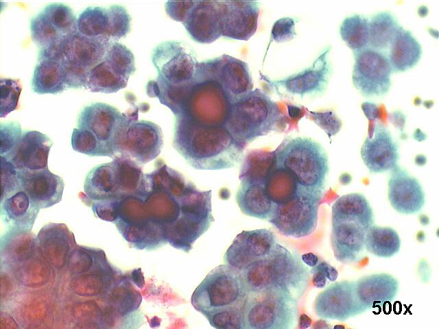
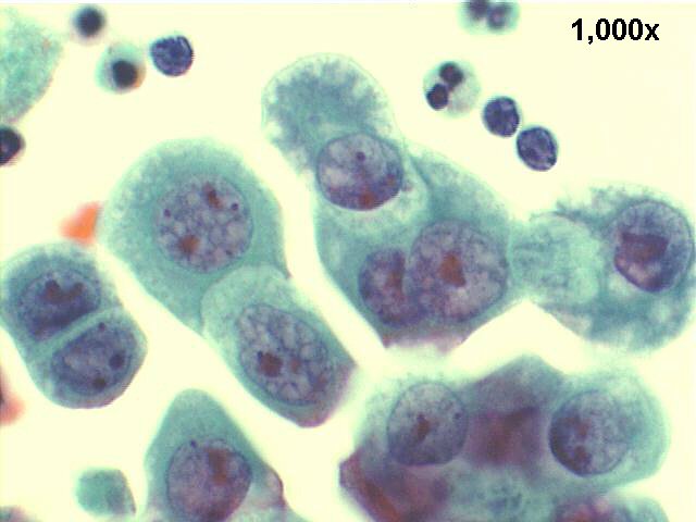
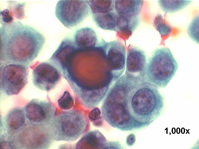
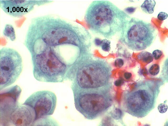
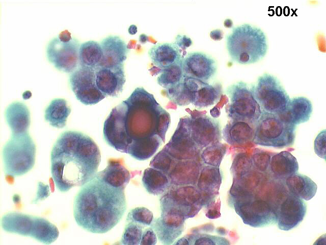
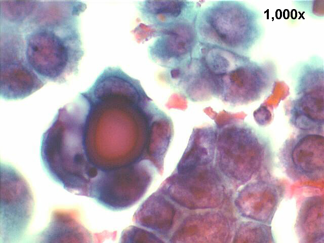
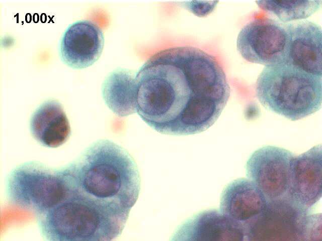
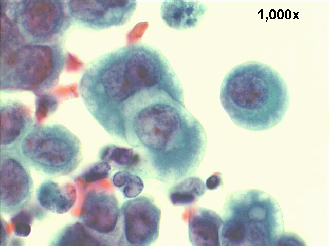
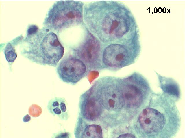
Ascites fluid, 73-year old female: papillary endometrial carcinoma


papillary carcinoma, endometrium,200x Pap staining, many acinar groups and psammoma bodies.

papillary carcinoma, endometrium,500x Pap staining, detail view of the psammoma bodies.

papillary carcinoma, endometrium,1000x Pap staining, slits between cells, and cells grasping other cells, suggestion of microvilli in their surface.

papillary carcinoma, endometrium, 1000x Pap staining, high power view of psammoma body, and cell inside cell arrangement.

papillary carcinoma, endometrium, 1000x Pap staining, slits between cells, and cells grasping other cells, suggestion of microvilli in their surface.

papillary carcinoma, endometrium, 500x Pap staining, acinar groups of cells, and psammoma body.

1000x Pap staining, high power view of previous picture.

papillary carcinoma, endometrium, 1000x Pap staining, high power view of cell grasping.

papillary carcinoma, endometrium, 1000x Pap staining, another high power view of cell grasping.

papillary carcinoma, endometrium, 1000x Pap staining, high power view of malignant cells in acinar arrangement. Notice very large and irregular nucleoli.


Adenocarcinoma, papillary, ascites fluid, primary in endometrium.

The smears show a typical papillary carcinoma, with massive number of acinar and papillary groups of cells, and many psammoma bodies. Very large cells with prominent nucleoli.


Features of Papillary Adenocarcinoma

Cellular patterns:
Three-dimensional groups, papillary arrangement, acinar, cell balls
Presence of psammoma bodies
Few single cells
Cytoplasm:
Basophilic, finely or coarsely vacuolated, cuboidal or round cells
Eccentrically placed nuclei, touching the cellular borders
Nucleus
Irregular chromatin pattern, with some coarse chromocenters
Prominent single or multiple nucleoli


Case A July 2007 References List of cases Atlas
Hosted by www.Geocities.ws

1