|
|
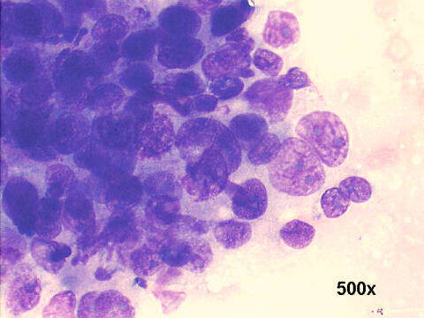
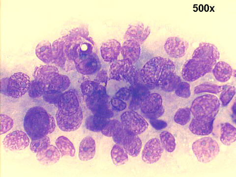
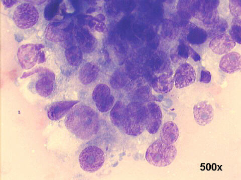
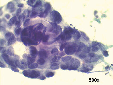
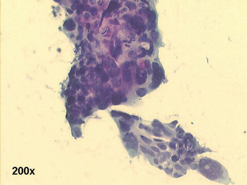
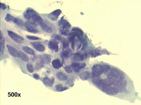
The smears show irregular tight clusters, overlapping nuclei, and few isolated malignant cells. The chromatin is moderately hyperchromatic, with some large nucleoli. These findings suggest recurrence of the previously treated poorly differentiated squamous cell carcinoma of the cervix. The imaging studies listed recurrence versus pelvic wall fibrosis due to radiotherapy as the main differential diagnosis, and the FNA provided the true diagnosis at minimal cost.

| Case A June 2003 | Case C June 2003 | References |
|
Atlas |