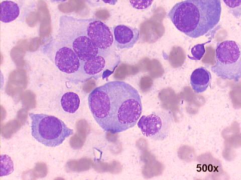
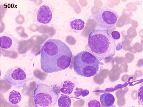
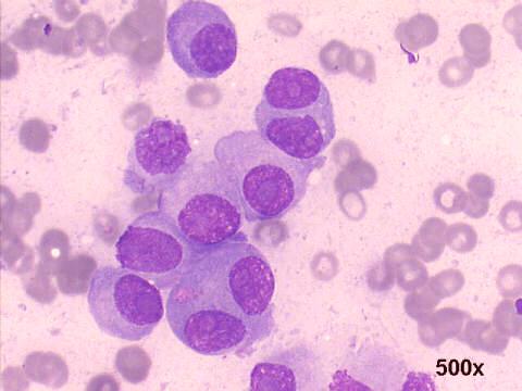
FNA thoracic mass: multiple myeloma
Drs. Prolla and Diehl's
INTERESTING CASE OF THE MONTH June 2002 Case 2
FNA cytology: many disperse "dyscohesive" malignant cells were seen, a quite specific pattern for malignancy. Many of them have the morphology of plasmablasts. Some have a curious and peculiar aspect, with two symmetrical nuclei with large nucleoli, reminding of E. T.'s head... At surgery, confirmation of the diagnosis of myelomatous lesion. Marker studies were positive only for kappa chains.