Drs. Prolla and Diehl's June 2006

Routine cervico-vaginal smear, 62-year old female: "blue blobs" in atrophic smear


100x Papanicolaou staining, demonstrating atrophic vaginitis with a background of basophilic granular debris.

200x Papanicolaou staining, demonstrating atrophic vaginitis with immature (parabasal) squamous epithelial cells with enlarged nuclei in a background of basophilic granular debris. Some round-shaped, amorphous basophilic structures ('blue blobs') are present.

200x Papanicolaou staining, demonstrating atrophic vaginitis with immature (parabasal) squamous epithelial cells with enlarged nuclei in a background of basophilic granular debris. Some round-shaped, amorphous basophilic structures ('blue blobs') are present.

500x Papanicolaou staining, demonstrating atrophic vaginitis with immature (parabasal) squamous epithelial cells with enlarged nuclei in a background of basophilic granular debris. Some round-shaped, amorphous basophilic structures ('blue blobs') are present.

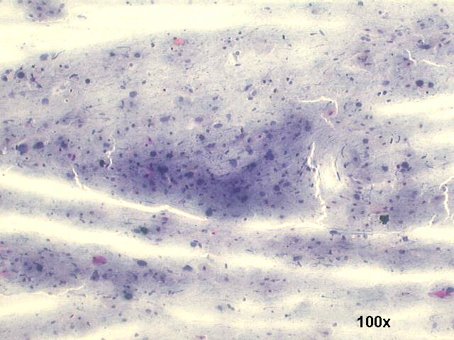
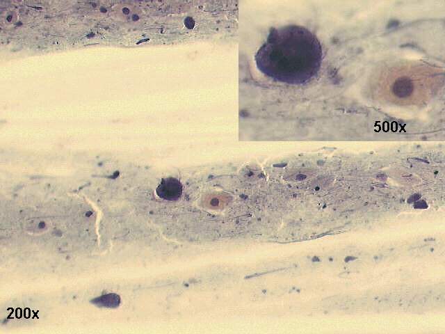
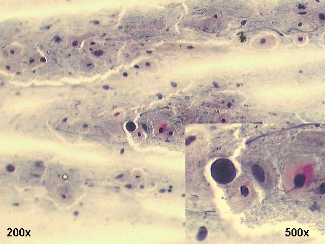
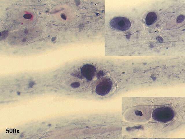
Dark blue rounded amorphous masses, known as "blue blobs", thought to represent degenerated nuclei or squamous cells can be seen in atrophic vaginal smears. In general, they appear to be of no clinical significance except as a source of potential diagnostic error, when mistaken for bare malignant nuclei.


Case June 2006 List of cases Atlas
Hosted by www.Geocities.ws

1