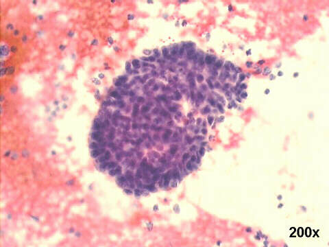
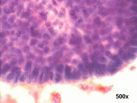
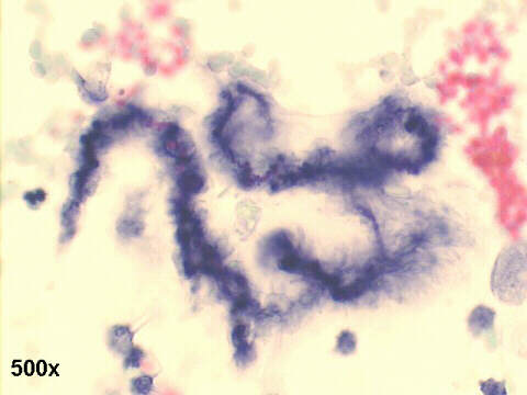
Ascites fluid cytopathology, 85-year old female, with known endometrial carcinoma.
|
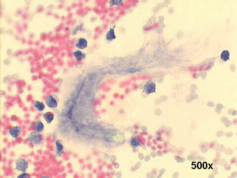
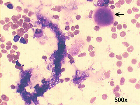
"Curschmann's spirals were found in smears and cell block preparations of five spontaneously occurring pleural and peritoneal fluids. The spirals were similar to those seen in the respiratory tract, although generally much smaller. In three of the five cases, the fluids also contained mucus-secreting adenocarcinoma cells; it is postulated that the spirals formed from mucus secreted by these cells. In the other two cases, there was evidence of serosal inflammation; it is suggested that the spirals in these cases developed from submesothelial connective tissue mucosubstances that entered the serosal cavity through a mesothelium of increased permeability due to the inflammation. No simple explanation can be accepted as to the exact mode of spiral formation, which is presumed to be a complex physical and biochemical phenomenon." |

| Back to Case June 2005 | References |
|
Atlas |