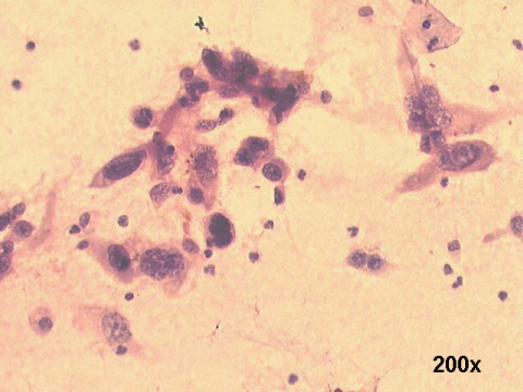
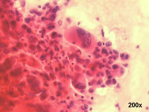
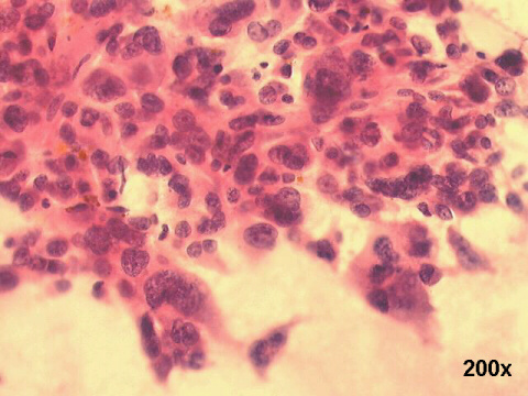
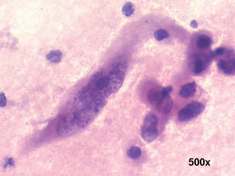
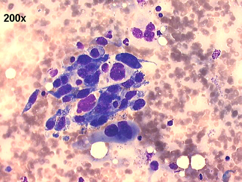
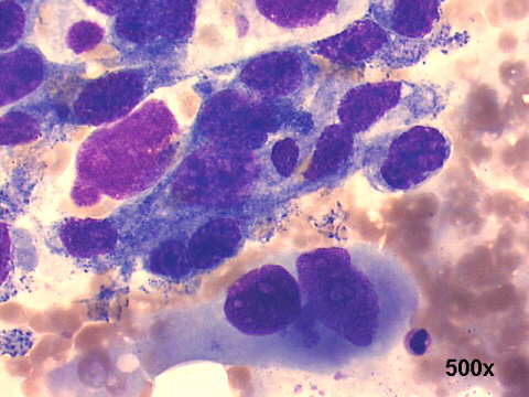
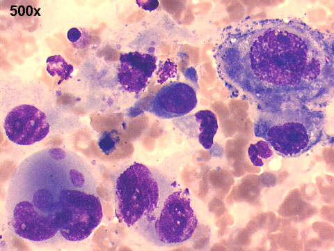
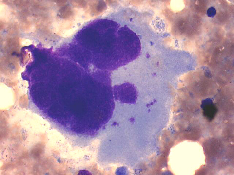
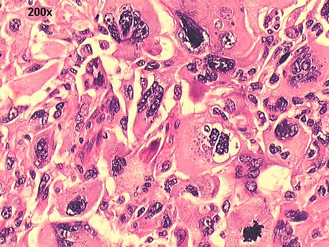
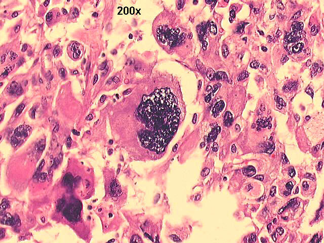
FNA right posterior costo-vertebral sulcus cytology, 68-year old male, 6-month post left lung transplantation: giant cell anaplastic carcinoma


Drs. Prolla and Diehl's INTERESTING CASE OF THE MONTH May 2004

200x Papanicolaou staining

200x Papanicolaou staining

200x Papanicolaou staining

500x Papanicolaou staining

200x M-G-G staining

500x M-G-G staining

500x M-G-G staining

500x M-G-G staining

200x HE staining

200x HE staining

The cytology smears show anaplastic malignant cells, some fusiform, some giant cells. Histology shows a giant cell anaplastic tumor, and immunocytochemistry was positive for epithelial origin markers, including the giant cells. This tumor is remarkable for several features: its extremely rapid growing, attaining 7cm in diameter six months after opposite lung transplantation, at which time high resolution CT of thorax did not reveal any tumor, and its giant cell epithelial nature.


References
List of cases
Atlas
Hosted by www.Geocities.ws

1