Pleural fluid: tuberculosis, lymphocytosis


Drs. Prolla and Diehl's INTERESTING CASE OF THE MONTH May 2003 case A

Tuberculosis, 500x Pap staining, normal looking lymphocytes are the predominant cells present

Tuberculosis, 500x Pap staining, normal looking lymphocytes are the predominant cells present, with very few epithelioid cells

500x Papanicolaou staining, 95% of cells are normal looking lymphocytes, and a Langhans type of histiocyte

200x M-G-G staining, 95% cells are normal looking lymphocytes

500x M-G-G staining, 95% lymphocytes

200x H&E staining, granulomatous pleuritis with Langhans histiocyte

500x H&E staining, detail of Langhans multinucleated histiocyte

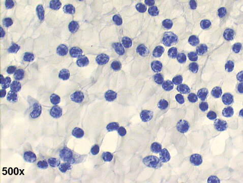
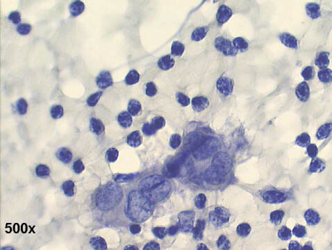
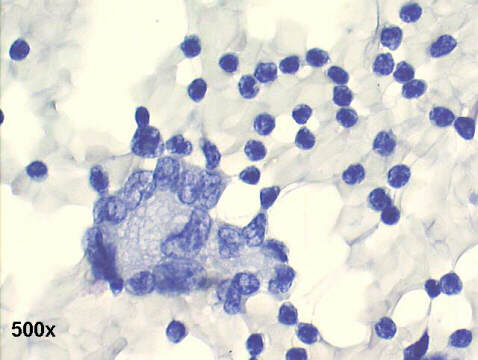
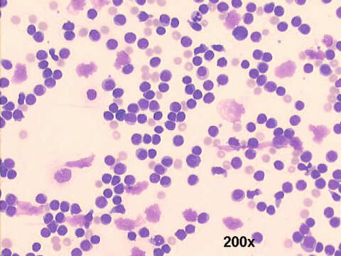
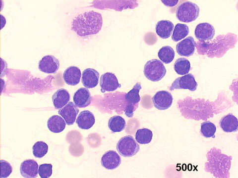
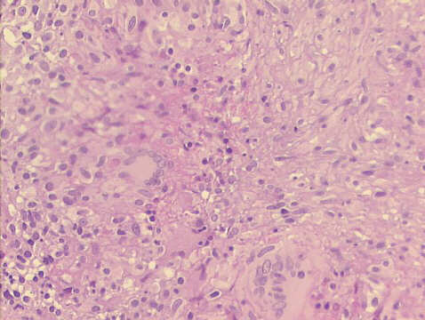
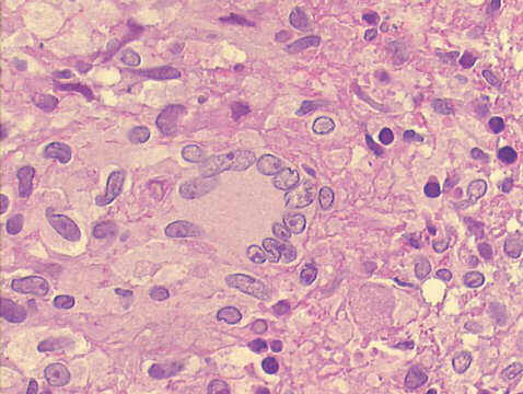
The pleural fluid sediment revealed the presence of many lymphocytes (approximately 95% of the cells present), some epithelioid cells, a few Langhans type of multinucleated histiocytes, and no mesothelial cells. A pleural biopsy showed granulomatous pleuritis, with positive Ziehl-Neelsen staining for acid fast bacilli. Cultures were positive for M. tuberculosis.
Lymphocytes in excess of 95% of the cellular content, and absence of mesothelial cells, are findings highly suggestive of tuberculosis in serous effusions. The presence of epithelioid cells, and Langhans cells are quite rare in cytological specimens of tuberculous pleuritis.


Case B May 2003 Case C May 2003 References
List of cases
Atlas
Hosted by www.Geocities.ws

1