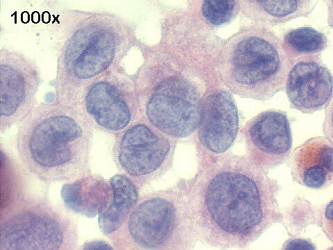
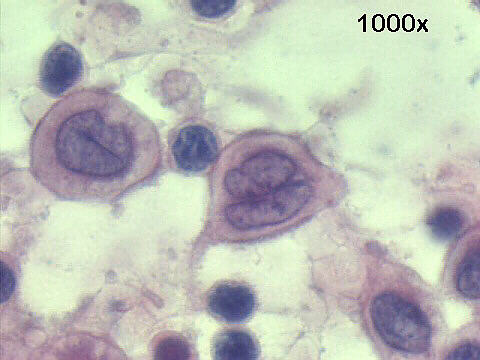
INTERESTING CASE OF THE MONTH - April 2000


FNA lymph node, Papanicolaou staining
Eosinophilic granuloma or Langerhans' cell histiocytosis

 Cytology of these cases is quite characteristic: very cellular smears, rich in Langerhans' histiocytes, eosinophils, some lymphocytes and plasma cells. Langerhans' histiocytes are medium to large cells, with characteristic nuclei: convoluted ones with longitudinal grooves. Most of them have a "coffee-bean" shape. Eosinophils are frequent  but not necessary for the diagnosis.


              Click here to see the histology of the case.


Features of Langherans´histiocytosis - Eosinophilic granuloma

Cellular patterns:
Large number of isolated or loosely aggregated Langherans´histiocytes
Variable number of eosinophils, plasma cells, lymphocytes
Variable number of multinucleated giant cells
Cytoplasm:
Abundant finely vacuolated cytoplasm
Nucleus
Folded or reniform nuclei, with longitudinal linear grooves
Few small or absent nucleoli
Finely granular chromatin pattern
Phenotypic Markers
Routinely express S-100, CD1a, OKT-6, HLA-DR markers
Birbeck´s granules or " zippers" at EM


Hospital de Clinicas de Porto Alegre, Pathology & Cytopathology Laboratories
Porto Alegre, RS Brazil 


Hospital de Clinicas de Porto Alegre - Listen to H.Bunji rendering of Mozart's Sonata in G K283


References
List of cases
Hosted by www.Geocities.ws

1