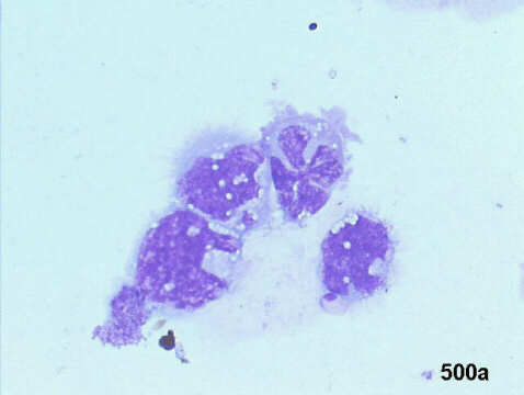
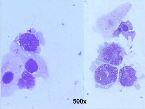
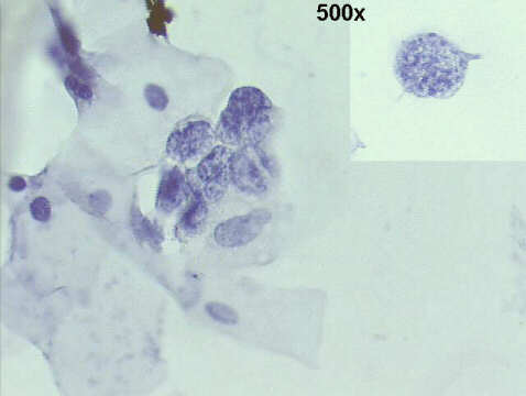
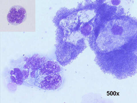
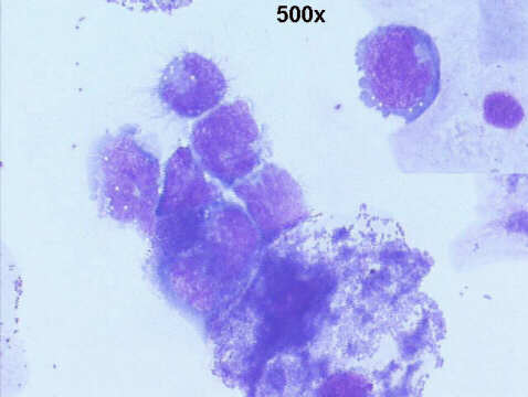
The patient had a known a B-cell lymphoma. The urine revealed the presence of many medium and large sized cleaved lymphoid cells, with a rim of basophilic cytoplasm, some with small lipid vacuoles, and a pattern of cytospin artifacts ("pseudo-flower cells"). Nuclei with moderately coarse chromatin.
These findings suggest the enlargement of the kidneys was due to lymphomatous infiltration. After several courses of chemotherapy, the kidneys reverted to normal size, and the urine was negative for lymphoma cells. This was not surprising, since with timely diagnosis and treatment, renal lesions may completely regress, often with minimal scarring of the renal parenchyma. The finding of lymphoma cells in urine is rarely reported, but this may be due to scarce use of urine cytology, even in the presence of presumed kidney involvement by lymphoma, rather than by low sensitivity.
Helical CT scans in this patient revealed bilateral nephromegaly, and a diffuse infiltrative pattern, with regular contour of the kidneys.
