|
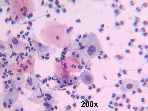
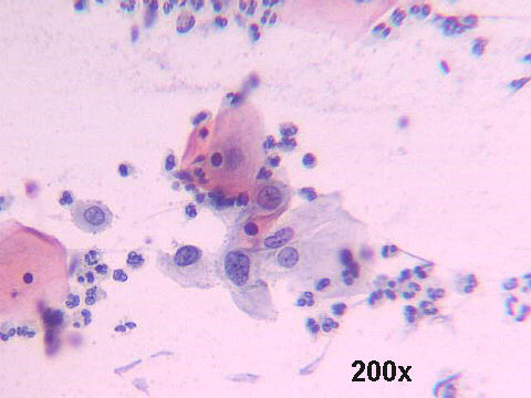
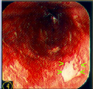 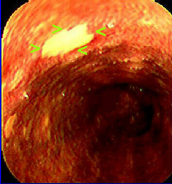
Ballon cytology: many "low grade" dysplastic squamous cells were seen. Increased N/C ratio, some binucleated cells, moderate nuclear hyperchromasia in other cells. At endoscopy, confirmation of the diagnosis, and identification of the dysplastic areas by negative lugol staining ( See article by Freitag et al. Dis Esophagus 1999;12:191-195.). Biopsy showed low grade intraepithelial squamous lesion. In Taquara, RS, Brazil, there is a high incidence of squamous cell carcinoma of the esophagus, and we are conducting a population surveillance with a special ballon for cell collection. This is one of the patients with dysplasia that were detected and treated (mucosectomy).
|